 Movie
Movie Controller


+ Open data

- Basic information
| Entry | Database: PDB / ID: 8twn | |||||||||||||||
|---|---|---|---|---|---|---|---|---|---|---|---|---|---|---|---|---|
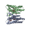
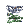
| Title | Crystal structure of nitrile synthase AetD with substrate bound | |||||||||||||||
 Components Components | AetD | |||||||||||||||
 Keywords Keywords | OXIDOREDUCTASE / nitrile synthase / non-heme iron enzyme / diiron enzyme | |||||||||||||||
| Function / homology | Haem oxygenase-like, multi-helical / metal ion binding / Chem-67I / D-MALATE / AetD Function and homology information Function and homology information | |||||||||||||||
| Biological species |  Aetokthonos hydrillicola (bacteria) Aetokthonos hydrillicola (bacteria) | |||||||||||||||
| Method |  X-RAY DIFFRACTION / SYNCHROTRON / SAD / Resolution: 2.08 Å X-RAY DIFFRACTION / SYNCHROTRON / SAD / Resolution: 2.08 Å | |||||||||||||||
 Authors Authors | Ye, N. / Drennan, C.L. | |||||||||||||||
| Funding support |  United States, 4items United States, 4items
| |||||||||||||||
 Citation Citation | Journal: Nat.Chem. / Year: 2024 Title: A single diiron enzyme catalyses the oxidative rearrangement of tryptophan to indole nitrile. Authors: Adak, S. / Ye, N. / Calderone, L.A. / Duan, M. / Lubeck, W. / Schafer, R.J.B. / Lukowski, A.L. / Houk, K.N. / Pandelia, M.E. / Drennan, C.L. / Moore, B.S. | |||||||||||||||
| History |
|
- Structure visualization
| Structure viewer | Molecule: MolmilJmol/JSmol |
|---|
- Downloads & links
-Download
| PDBx/mmCIF format | 8twn.cif.gz | 77.1 KB | Display | PDBx/mmCIF format |
|---|---|---|---|---|
| PDB format | pdb8twn.ent.gz | 48 KB | Display | PDB format |
| PDBx/mmJSON format | 8twn.json.gz | Tree view | PDBx/mmJSON format | |
| Others |  Other downloads Other downloads |
-Validation report
| Arichive directory | https://data.pdbj.org/pub/pdb/validation_reports/tw/8twnftp://data.pdbj.org/pub/pdb/validation_reports/tw/8twn | HTTPS FTP |
|---|
-Related structure data
-Links
 PDBj
PDBj

- Assembly
| Deposited unit | 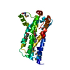
| ||||||||||||
|---|---|---|---|---|---|---|---|---|---|---|---|---|---|
| 1 |
| ||||||||||||
| Unit cell |
|
-Components
| #1: Protein | Mass: 30640.727 Da / Num. of mol.: 1 Source method: isolated from a genetically manipulated source Source: (gene. exp.) Aetokthonos hydrillicola (bacteria) / Gene: aetD / Production host: |
|---|---|
| #2: Chemical | ChemComp-67I / (  Mass: 362.017 Da / Num. of mol.: 1 / Source method: obtained synthetically / Formula: C11H10Br2N2O2 / Feature type: SUBJECT OF INVESTIGATION Mass: 362.017 Da / Num. of mol.: 1 / Source method: obtained synthetically / Formula: C11H10Br2N2O2 / Feature type: SUBJECT OF INVESTIGATION |
| #3: Chemical | ChemComp-MLT /   Mass: 134.087 Da / Num. of mol.: 1 / Source method: obtained synthetically / Formula: C4H6O5 Mass: 134.087 Da / Num. of mol.: 1 / Source method: obtained synthetically / Formula: C4H6O5 |
| #4: Water | ChemComp-HOH /  Mass: 18.015 Da / Num. of mol.: 98 / Source method: isolated from a natural source / Formula: H2O Mass: 18.015 Da / Num. of mol.: 98 / Source method: isolated from a natural source / Formula: H2O |
| Has ligand of interest | Y |
| Has protein modification | Y |
-Experimental details
-Experiment
| Experiment | Method: X-RAY DIFFRACTION / Number of used crystals: 1 |
|---|
- Sample preparation
| Crystal | Density Matthews: 3.14 Å3/Da / Density % sol: 60.78 % |
|---|---|
| Crystal grow | Temperature: 295 K / Method: vapor diffusion, hanging drop / pH: 6 Details: 100 mM MES monohydrate pH 6.0, 20% PEG 4000, 110 mM malonic acid, 15 mM ammonium citrate tribasic, 7.2 mM succinic acid, 18 mM DL-malic acid, 24 mM sodium acetate trihydrate, 30 mM sodium ...Details: 100 mM MES monohydrate pH 6.0, 20% PEG 4000, 110 mM malonic acid, 15 mM ammonium citrate tribasic, 7.2 mM succinic acid, 18 mM DL-malic acid, 24 mM sodium acetate trihydrate, 30 mM sodium formate, 9.6 mM ammonium tartrate dibasic |
-Data collection
| Diffraction | Mean temperature: 100 K / Serial crystal experiment: N |
|---|---|
| Diffraction source | Source: SYNCHROTRON / Site: APS / Beamline: 24-ID-C / Wavelength: 0.9792 Å |
| Detector | Type: DECTRIS EIGER2 X 16M / Detector: PIXEL / Date: Jul 20, 2022 |
| Radiation | Monochromator: Double crystal Si(111) / Protocol: SINGLE WAVELENGTH / Monochromatic (M) / Laue (L): M / Scattering type: x-ray |
| Radiation wavelength | Wavelength: 0.9792 Å / Relative weight: 1 |
| Reflection | Resolution: 2.08→50 Å / Num. obs: 22444 / % possible obs: 92.4 % / Redundancy: 9.1 % / Biso Wilson estimate: 42.89 Å2 / CC1/2: 0.995 / Net I/σ(I): 9.6 |
| Reflection shell | Resolution: 2.08→2.21 Å / Num. unique obs: 3470 / CC1/2: 0.844 |
- Processing
| Software |
| |||||||||||||||||||||||||||||||||||||||||||||||||||||||||||||||
|---|---|---|---|---|---|---|---|---|---|---|---|---|---|---|---|---|---|---|---|---|---|---|---|---|---|---|---|---|---|---|---|---|---|---|---|---|---|---|---|---|---|---|---|---|---|---|---|---|---|---|---|---|---|---|---|---|---|---|---|---|---|---|---|---|
| Refinement | Method to determine structure: SAD / Resolution: 2.08→47.25 Å / SU ML: 0.2551 / Cross valid method: FREE R-VALUE / σ(F): 1.35 / Phase error: 22.045 Stereochemistry target values: GeoStd + Monomer Library + CDL v1.2
| |||||||||||||||||||||||||||||||||||||||||||||||||||||||||||||||
| Solvent computation | Shrinkage radii: 0.9 Å / VDW probe radii: 1.1 Å / Solvent model: FLAT BULK SOLVENT MODEL | |||||||||||||||||||||||||||||||||||||||||||||||||||||||||||||||
| Displacement parameters | Biso mean: 45.75 Å2 | |||||||||||||||||||||||||||||||||||||||||||||||||||||||||||||||
| Refinement step | Cycle: LAST / Resolution: 2.08→47.25 Å
| |||||||||||||||||||||||||||||||||||||||||||||||||||||||||||||||
| Refine LS restraints |
| |||||||||||||||||||||||||||||||||||||||||||||||||||||||||||||||
| LS refinement shell |
|
