 Movie
Movie Controller


+ Open data

- Basic information
| Entry | Database: PDB / ID: 8tu9 | ||||||
|---|---|---|---|---|---|---|---|
| Title | Cryo-EM structure of HGSNAT-acetyl-CoA complex at pH 7.5 | ||||||
 Components Components | Enhanced green fluorescent protein,Heparan-alpha-glucosaminide N-acetyltransferase,Isoform 2 of Heparan-alpha-glucosaminide N-acetyltransferase | ||||||
 Keywords Keywords | TRANSFERASE / Heparan-alpha-glucosaminide N-acetyltransferase / Transmembrane protein 76 / membrane protein | ||||||
| Function / homology |  Function and homology information Function and homology informationheparan-alpha-glucosaminide N-acetyltransferase / heparan-alpha-glucosaminide N-acetyltransferase activity / MPS IIIC - Sanfilippo syndrome C / heparan sulfate proteoglycan catabolic process / HS-GAG degradation / lysosomal transport / acyltransferase activity / protein complex oligomerization / tertiary granule membrane / specific granule membrane ...heparan-alpha-glucosaminide N-acetyltransferase / heparan-alpha-glucosaminide N-acetyltransferase activity / MPS IIIC - Sanfilippo syndrome C / heparan sulfate proteoglycan catabolic process / HS-GAG degradation / lysosomal transport / acyltransferase activity / protein complex oligomerization / tertiary granule membrane / specific granule membrane / lysosomal lumen / bioluminescence / generation of precursor metabolites and energy / lysosomal membrane / Neutrophil degranulation / plasma membrane Similarity search - Function | ||||||
| Biological species |   Aequorea victoria (jellyfish) Aequorea victoria (jellyfish) Homo sapiens (human) Homo sapiens (human) | ||||||
| Method | ELECTRON MICROSCOPY / single particle reconstruction / cryo EM / Resolution: 3.26 Å | ||||||
 Authors Authors | Navratna, V. / Kumar, A. / Mosalaganti, S. | ||||||
| Funding support | 1items
| ||||||
 Citation Citation | Journal: Elife / Year: 2024 Title: Structure of the human heparan-α-glucosaminide -acetyltransferase (HGSNAT). Authors: Vikas Navratna / Arvind Kumar / Jaimin K Rana / Shyamal Mosalaganti /  Abstract: Degradation of heparan sulfate (HS), a glycosaminoglycan (GAG) comprised of repeating units of -acetylglucosamine and glucuronic acid, begins in the cytosol and is completed in the lysosomes. ...Degradation of heparan sulfate (HS), a glycosaminoglycan (GAG) comprised of repeating units of -acetylglucosamine and glucuronic acid, begins in the cytosol and is completed in the lysosomes. Acetylation of the terminal non-reducing amino group of α-D-glucosamine of HS is essential for its complete breakdown into monosaccharides and free sulfate. Heparan-α-glucosaminide -acetyltransferase (HGSNAT), a resident of the lysosomal membrane, catalyzes this essential acetylation reaction by accepting and transferring the acetyl group from cytosolic acetyl-CoA to terminal α-D-glucosamine of HS in the lysosomal lumen. Mutation-induced dysfunction in HGSNAT causes abnormal accumulation of HS within the lysosomes and leads to an autosomal recessive neurodegenerative lysosomal storage disorder called mucopolysaccharidosis IIIC (MPS IIIC). There are no approved drugs or treatment strategies to cure or manage the symptoms of, MPS IIIC. Here, we use cryo-electron microscopy (cryo-EM) to determine a high-resolution structure of the HGSNAT-acetyl-CoA complex, the first step in the HGSNAT-catalyzed acetyltransferase reaction. In addition, we map the known MPS IIIC mutations onto the structure and elucidate the molecular basis for mutation-induced HGSNAT dysfunction. #1: Journal: eLife / Year: 2024Title: Structure of the human heparan-alpha-glucosaminide N-acetyltransferase (HGSNAT) Authors: Navratna, V. / Kumar, A. / Mosalaganti, S. #2: Journal: bioRxiv / Year: 2024 Title: Structure of the human heparan-α-glucosaminide -acetyltransferase (HGSNAT). Authors: Vikas Navratna / Arvind Kumar / Jaimin K Rana / Shyamal Mosalaganti / Abstract: Degradation of heparan sulfate (HS), a glycosaminoglycan (GAG) comprised of repeating units of -acetylglucosamine and glucuronic acid, begins in the cytosol and is completed in the lysosomes. ...Degradation of heparan sulfate (HS), a glycosaminoglycan (GAG) comprised of repeating units of -acetylglucosamine and glucuronic acid, begins in the cytosol and is completed in the lysosomes. Acetylation of the terminal non-reducing amino group of a-D-glucosamine of HS is essential for its complete breakdown into monosaccharides and free sulfate. Heparan-a-glucosaminide -acetyltransferase (HGSNAT), a resident of the lysosomal membrane, catalyzes this essential acetylation reaction by accepting and transferring the acetyl group from cytosolic acetyl-CoA to terminal a-D-glucosamine of HS in the lysosomal lumen. Mutation-induced dysfunction in HGSNAT causes abnormal accumulation of HS within the lysosomes and leads to an autosomal recessive neurodegenerative lysosomal storage disorder called mucopolysaccharidosis IIIC (MPS IIIC). There are no approved drugs or treatment strategies to cure or manage the symptoms of, MPS IIIC. Here, we use cryo-electron microscopy (cryo-EM) to determine a high-resolution structure of the HGSNAT-acetyl-CoA complex, the first step in HGSNAT catalyzed acetyltransferase reaction. In addition, we map the known MPS IIIC mutations onto the structure and elucidate the molecular basis for mutation-induced HGSNAT dysfunction. | ||||||
| History |
|
- Structure visualization
| Structure viewer | Molecule: MolmilJmol/JSmol |
|---|
- Downloads & links
-Download
| PDBx/mmCIF format | 8tu9.cif.gz | 214.5 KB | Display | PDBx/mmCIF format |
|---|---|---|---|---|
| PDB format | pdb8tu9.ent.gz | 158.5 KB | Display | PDB format |
| PDBx/mmJSON format | 8tu9.json.gz | Tree view | PDBx/mmJSON format | |
| Others |  Other downloads Other downloads |
-Validation report
| Summary document | 8tu9_validation.pdf.gz | 1.7 MB | Display | wwPDB validaton report |
|---|---|---|---|---|
| Full document | 8tu9_full_validation.pdf.gz | 1.8 MB | Display | |
| Data in XML | 8tu9_validation.xml.gz | 48.6 KB | Display | |
| Data in CIF | 8tu9_validation.cif.gz | 70.8 KB | Display | |
| Arichive directory | https://data.pdbj.org/pub/pdb/validation_reports/tu/8tu9ftp://data.pdbj.org/pub/pdb/validation_reports/tu/8tu9 | HTTPS FTP |
-Related structure data
| Related structure data |  41620MC M: map data used to model this data C: citing same article ( |
|---|---|
| Similar structure data |
-Links
 PDBj
PDBj

- Assembly
| Deposited unit | 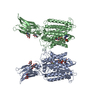
| |||||||||||||||||||||||||||||||||||||||||||||||||||||||||||
|---|---|---|---|---|---|---|---|---|---|---|---|---|---|---|---|---|---|---|---|---|---|---|---|---|---|---|---|---|---|---|---|---|---|---|---|---|---|---|---|---|---|---|---|---|---|---|---|---|---|---|---|---|---|---|---|---|---|---|---|---|
| 1 |
| |||||||||||||||||||||||||||||||||||||||||||||||||||||||||||
| Noncrystallographic symmetry (NCS) | NCS domain:
NCS domain segments: Ens-ID: ens_1
NCS oper: (Code: givenMatrix: (-0.999999634014, -0.000836221116112, 0.000180850914369), (0.00083627082141, -0.999999612529, 0.000274940524232), (0.000180620933222, 0.00027509166395, 0.99999994585) ...NCS oper: (Code: given Matrix: (-0.999999634014, -0.000836221116112, 0.000180850914369), Vector: |
-Components
| #1: Protein | Mass: 100817.172 Da / Num. of mol.: 2 Source method: isolated from a genetically manipulated source Source: (gene. exp.) Aequorea victoria (jellyfish), (gene. exp.) Homo sapiens (human)Gene: MMB69_26960, HGSNAT, TMEM76 / Plasmid: pEG BacMam N term StrepII eGFP 3C / Details (production host): Addgene # 160683 / Cell line (production host): HEK293S GnTI- / Production host: Homo sapiens (human)References: UniProt: A0A9X4KGN5, UniProt: Q68CP4, heparan-alpha-glucosaminide N-acetyltransferase #2: Chemical |   Mass: 809.571 Da / Num. of mol.: 2 / Source method: obtained synthetically / Formula: C23H38N7O17P3S / Feature type: SUBJECT OF INVESTIGATION Mass: 809.571 Da / Num. of mol.: 2 / Source method: obtained synthetically / Formula: C23H38N7O17P3S / Feature type: SUBJECT OF INVESTIGATION#3: Sugar | ChemComp-NAG /   Type: D-saccharide, beta linking / Mass: 221.208 Da / Num. of mol.: 6 / Source method: obtained synthetically / Formula: C8H15NO6 / Feature type: SUBJECT OF INVESTIGATION Type: D-saccharide, beta linking / Mass: 221.208 Da / Num. of mol.: 6 / Source method: obtained synthetically / Formula: C8H15NO6 / Feature type: SUBJECT OF INVESTIGATIONHas ligand of interest | Y | Has protein modification | Y | |
|---|
-Experimental details
-Experiment
| Experiment | Method: ELECTRON MICROSCOPY |
|---|---|
| EM experiment | Aggregation state: PARTICLE / 3D reconstruction method: single particle reconstruction |
- Sample preparation
| Component | Name: Heparan acetyl-CoA: alpha-glucosaminide N-acetyltransferase (HGSNAT) Type: COMPLEX Details: Full length (Isoform 2) human HGSNAT expressed as a recombinant fusion protein with N-terminal GFP, in mammalian cells. Entity ID: #1 / Source: MULTIPLE SOURCES | ||||||||||||||||||||
|---|---|---|---|---|---|---|---|---|---|---|---|---|---|---|---|---|---|---|---|---|---|
| Molecular weight | Value: 0.1007 MDa / Experimental value: NO | ||||||||||||||||||||
| Source (natural) |
| ||||||||||||||||||||
| Source (recombinant) | Organism: Homo sapiens (human) / Cell: HEK293S GnTI- / Plasmid: pEG BacMam N term StrepII eGFP 3C | ||||||||||||||||||||
| Buffer solution | pH: 7.5 | ||||||||||||||||||||
| Buffer component |
| ||||||||||||||||||||
| Specimen | Conc.: 0.9 mg/ml / Embedding applied: NO / Shadowing applied: NO / Staining applied: NO / Vitrification applied: YES Details: HGSNAT in digitonin micelle, purified by Strep-Tactin affinity chromatography. | ||||||||||||||||||||
| Specimen support | Details: 15 mA current / Grid material: GOLD / Grid mesh size: 300 divisions/in. / Grid type: UltrAuFoil R1.2/1.3 | ||||||||||||||||||||
| Vitrification | Instrument: FEI VITROBOT MARK IV / Cryogen name: ETHANE / Humidity: 100 % / Chamber temperature: 291 K |
- Electron microscopy imaging
| Experimental equipment |  Model: Titan Krios / Image courtesy: FEI Company |
|---|---|
| Microscopy | Model: FEI TITAN KRIOS |
| Electron gun | Electron source:  FIELD EMISSION GUN / Accelerating voltage: 300 kV / Illumination mode: FLOOD BEAM FIELD EMISSION GUN / Accelerating voltage: 300 kV / Illumination mode: FLOOD BEAM |
| Electron lens | Mode: BRIGHT FIELD / Nominal defocus max: 2500 nm / Nominal defocus min: 1000 nm / Cs: 2.7 mm / C2 aperture diameter: 100 µm |
| Specimen holder | Cryogen: NITROGEN / Specimen holder model: FEI TITAN KRIOS AUTOGRID HOLDER |
| Image recording | Average exposure time: 2 sec. / Electron dose: 50 e/Å2 / Film or detector model: GATAN K3 BIOQUANTUM (6k x 4k) / Num. of grids imaged: 2 / Num. of real images: 10000 |
| Image scans | Width: 5760 / Height: 4092 |
- Processing
| EM software |
| ||||||||||||||||||||||||
|---|---|---|---|---|---|---|---|---|---|---|---|---|---|---|---|---|---|---|---|---|---|---|---|---|---|
| CTF correction | Type: PHASE FLIPPING AND AMPLITUDE CORRECTION | ||||||||||||||||||||||||
| Particle selection | Num. of particles selected: 15000000 | ||||||||||||||||||||||||
| Symmetry | Point symmetry: C2 (2 fold cyclic) | ||||||||||||||||||||||||
| 3D reconstruction | Resolution: 3.26 Å / Resolution method: FSC 0.143 CUT-OFF / Num. of particles: 57739 / Symmetry type: POINT | ||||||||||||||||||||||||
| Atomic model building | Protocol: RIGID BODY FIT | ||||||||||||||||||||||||
| Atomic model building | Details: Made using Model angelo / Source name: Other / Type: in silico model | ||||||||||||||||||||||||
| Refinement | Cross valid method: NONE Stereochemistry target values: GeoStd + Monomer Library + CDL v1.2 | ||||||||||||||||||||||||
| Displacement parameters | Biso mean: 20.21 Å2 | ||||||||||||||||||||||||
| Refine LS restraints |
| ||||||||||||||||||||||||
| Refine LS restraints NCS | Type: NCS constraints / Rms dev position: 8.33142807632E-11 Å |
