 Movie
Movie Controller


+ Open data

- Basic information
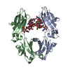
| Entry | Database: PDB / ID: 8ttm | ||||||
|---|---|---|---|---|---|---|---|
| Title | IgG1 Fc Heterodimer combYSelect1 | ||||||
 Components Components | (Immunoglobulin gamma-1 heavy chain) x 2 | ||||||
 Keywords Keywords | IMMUNE SYSTEM / IgG1 Fc / heterodimer | ||||||
| Function / homology |  Function and homology information Function and homology informationimmunoglobulin complex / adaptive immune response / extracellular region / plasma membrane Similarity search - Function | ||||||
| Biological species |  Homo sapiens x Mus musculus hybrid cell line (mammal) Homo sapiens x Mus musculus hybrid cell line (mammal) | ||||||
| Method |  X-RAY DIFFRACTION / SYNCHROTRON / MOLECULAR REPLACEMENT / Resolution: 2.51 Å X-RAY DIFFRACTION / SYNCHROTRON / MOLECULAR REPLACEMENT / Resolution: 2.51 Å | ||||||
 Authors Authors | Azzam, T. / Du, J.J. / Sundberg, E.J. | ||||||
| Funding support |  United States, 1items United States, 1items
| ||||||
 Citation Citation | Journal: Sci Adv / Year: 2024 Title: Combinatorially restricted computational design of protein-protein interfaces to produce IgG heterodimers. Authors: Azzam, T. / Du, J.J. / Flowers, M.W. / Ali, A.V. / Hunn, J.C. / Vijayvargiya, N. / Knagaram, R. / Bogacz, M. / Maravillas, K.E. / Sastre, D.E. / Fields, J.K. / Mirzaei, A. / Pierce, B.G. / Sundberg, E.J. | ||||||
| History |
|
- Structure visualization
| Structure viewer | Molecule: MolmilJmol/JSmol |
|---|
- Downloads & links
-Download
| PDBx/mmCIF format | 8ttm.cif.gz | 190.7 KB | Display | PDBx/mmCIF format |
|---|---|---|---|---|
| PDB format | pdb8ttm.ent.gz | 150.8 KB | Display | PDB format |
| PDBx/mmJSON format | 8ttm.json.gz | Tree view | PDBx/mmJSON format | |
| Others |  Other downloads Other downloads |
-Validation report
| Summary document | 8ttm_validation.pdf.gz | 1.2 MB | Display | wwPDB validaton report |
|---|---|---|---|---|
| Full document | 8ttm_full_validation.pdf.gz | 1.2 MB | Display | |
| Data in XML | 8ttm_validation.xml.gz | 17.9 KB | Display | |
| Data in CIF | 8ttm_validation.cif.gz | 23.6 KB | Display | |
| Arichive directory | https://data.pdbj.org/pub/pdb/validation_reports/tt/8ttmftp://data.pdbj.org/pub/pdb/validation_reports/tt/8ttm | HTTPS FTP |
-Related structure data
-Links
 PDBj
PDBj


- Assembly
| Deposited unit | 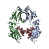
| ||||||||
|---|---|---|---|---|---|---|---|---|---|
| 1 |
| ||||||||
| Unit cell |
|
-Components
| #1: Antibody | Mass: 26146.555 Da / Num. of mol.: 1 / Mutation: K409S, T411Y Source method: isolated from a genetically manipulated source Source: (gene. exp.) Homo sapiens x Mus musculus hybrid cell line (mammal)Production host:  Homo sapiens (human) / References: UniProt: P0DOX5 Homo sapiens (human) / References: UniProt: P0DOX5 | ||||||
|---|---|---|---|---|---|---|---|
| #2: Antibody | Mass: 26148.592 Da / Num. of mol.: 1 / Mutation: L368S, D399Y Source method: isolated from a genetically manipulated source Source: (gene. exp.) Homo sapiens x Mus musculus hybrid cell line (mammal)Production host: Homo sapiens (human) / References: UniProt: P0DOX5 | ||||||
| #3: Polysaccharide | Type: oligosaccharide / Mass: 1463.349 Da / Num. of mol.: 2 Source method: isolated from a genetically manipulated source #4: Water | ChemComp-HOH / |  Mass: 18.015 Da / Num. of mol.: 6 / Source method: isolated from a natural source / Formula: H2O Mass: 18.015 Da / Num. of mol.: 6 / Source method: isolated from a natural source / Formula: H2OHas ligand of interest | N | Has protein modification | Y | |
-Experimental details
-Experiment
| Experiment | Method: X-RAY DIFFRACTION / Number of used crystals: 1 |
|---|
- Sample preparation
| Crystal | Density Matthews: 2.7 Å3/Da / Density % sol: 54.45 % |
|---|---|
| Crystal grow | Temperature: 292 K / Method: vapor diffusion, sitting drop / pH: 6.5 / Details: 0.1M BisTris pH 6.5 and 21% PEG MME 5000 |
-Data collection
| Diffraction | Mean temperature: 100 K / Serial crystal experiment: N |
|---|---|
| Diffraction source | Source: SYNCHROTRON / Site: APS / Beamline: 22-ID / Wavelength: 1 Å |
| Detector | Type: DECTRIS EIGER X 16M / Detector: PIXEL / Date: Mar 4, 2023 |
| Radiation | Protocol: SINGLE WAVELENGTH / Monochromatic (M) / Laue (L): M / Scattering type: x-ray |
| Radiation wavelength | Wavelength: 1 Å / Relative weight: 1 |
| Reflection | Resolution: 2.51→40.88 Å / Num. obs: 19700 / % possible obs: 98.13 % / Redundancy: 10.2 % / CC1/2: 0.993 / Rmerge(I) obs: 0.24 / Rpim(I) all: 0.079 / Rrim(I) all: 0.254 / Net I/σ(I): 30.3 |
| Reflection shell | Resolution: 2.51→2.6 Å / Rmerge(I) obs: 0.961 / Mean I/σ(I) obs: 2.25 / Num. unique obs: 1814 / CC1/2: 0.852 / Rpim(I) all: 0.336 / Rrim(I) all: 1.02 |
- Processing
| Software |
| |||||||||||||||||||||||||||||||||||||||||||||||||||||||||||||||||||||||||||||||||||||||||||||||
|---|---|---|---|---|---|---|---|---|---|---|---|---|---|---|---|---|---|---|---|---|---|---|---|---|---|---|---|---|---|---|---|---|---|---|---|---|---|---|---|---|---|---|---|---|---|---|---|---|---|---|---|---|---|---|---|---|---|---|---|---|---|---|---|---|---|---|---|---|---|---|---|---|---|---|---|---|---|---|---|---|---|---|---|---|---|---|---|---|---|---|---|---|---|---|---|---|
| Refinement | Method to determine structure: MOLECULAR REPLACEMENT / Resolution: 2.51→40.88 Å / Cor.coef. Fo:Fc: 0.955 / Cor.coef. Fo:Fc free: 0.937 / SU B: 21.273 / SU ML: 0.213 / Cross valid method: THROUGHOUT / ESU R: 0.529 / ESU R Free: 0.284 / Stereochemistry target values: MAXIMUM LIKELIHOOD Details: U VALUES : WITH TLS ADDED HYDROGENS HAVE BEEN ADDED IN THE RIDING POSITIONS U VALUES : RESIDUAL ONLY
| |||||||||||||||||||||||||||||||||||||||||||||||||||||||||||||||||||||||||||||||||||||||||||||||
| Solvent computation | Ion probe radii: 0.7 Å / Shrinkage radii: 0.7 Å / VDW probe radii: 1 Å / Solvent model: MASK | |||||||||||||||||||||||||||||||||||||||||||||||||||||||||||||||||||||||||||||||||||||||||||||||
| Displacement parameters | Biso mean: 77.516 Å2
| |||||||||||||||||||||||||||||||||||||||||||||||||||||||||||||||||||||||||||||||||||||||||||||||
| Refinement step | Cycle: LAST / Resolution: 2.51→40.88 Å
| |||||||||||||||||||||||||||||||||||||||||||||||||||||||||||||||||||||||||||||||||||||||||||||||
| Refine LS restraints |
| |||||||||||||||||||||||||||||||||||||||||||||||||||||||||||||||||||||||||||||||||||||||||||||||
| LS refinement shell | Resolution: 2.51→2.575 Å / Total num. of bins used: 20
| |||||||||||||||||||||||||||||||||||||||||||||||||||||||||||||||||||||||||||||||||||||||||||||||
| Refinement TLS params. | Method: refined / Refine-ID: X-RAY DIFFRACTION
| |||||||||||||||||||||||||||||||||||||||||||||||||||||||||||||||||||||||||||||||||||||||||||||||
| Refinement TLS group |
|
