 Movie
Movie Controller


+ Open data

- Basic information
| Entry | Database: PDB / ID: 8tsm | ||||||
|---|---|---|---|---|---|---|---|
| Title | Crystal structure of chicken Netrin-1 LN LE1-2 | ||||||
 Components Components | Netrin-1 | ||||||
 Keywords Keywords | SIGNALING PROTEIN / Extracellular matrix protein / apoptosis / axon guidance / neuronal development / cell signaling / heparan sulfate binding protein / dependence receptors | ||||||
| Function / homology |  Function and homology information Function and homology informationregulation of glial cell migration / chemorepulsion of axon / Cdc42 protein signal transduction / anterior/posterior axon guidance / motor neuron migration / negative regulation of axon extension / substrate-dependent cell migration, cell extension / positive regulation of cell motility / inner ear morphogenesis / nuclear migration ...regulation of glial cell migration / chemorepulsion of axon / Cdc42 protein signal transduction / anterior/posterior axon guidance / motor neuron migration / negative regulation of axon extension / substrate-dependent cell migration, cell extension / positive regulation of cell motility / inner ear morphogenesis / nuclear migration / regulation of synapse assembly / basement membrane / positive regulation of glial cell proliferation / positive regulation of axon extension / glial cell proliferation / cell periphery / cell-cell adhesion / actin cytoskeleton / Ras protein signal transduction / glutamatergic synapse / extracellular region / nucleoplasm / cytosol Similarity search - Function | ||||||
| Biological species |  | ||||||
| Method |  X-RAY DIFFRACTION / SYNCHROTRON / MOLECULAR REPLACEMENT / Resolution: 3.9 Å X-RAY DIFFRACTION / SYNCHROTRON / MOLECULAR REPLACEMENT / Resolution: 3.9 Å | ||||||
 Authors Authors | Heide, F. / Rafiei, F. / Stetefeld, J. | ||||||
| Funding support |  Canada, 1items Canada, 1items
| ||||||
 Citation Citation | Journal: To Be Published Title: Investigation of the dynamic nature of Netrin-1 on dependence receptor signaling Authors: Rafiei, F. / Heide, F. / Legare, S. / Gabir, H. / Padilla-Meier, P. / Meier, M. / Koch, M. / Stetefeld, J. | ||||||
| History |
|
- Structure visualization
| Structure viewer | Molecule: MolmilJmol/JSmol |
|---|
- Downloads & links
-Download
| PDBx/mmCIF format | 8tsm.cif.gz | 257 KB | Display | PDBx/mmCIF format |
|---|---|---|---|---|
| PDB format | pdb8tsm.ent.gz | 178.3 KB | Display | PDB format |
| PDBx/mmJSON format | 8tsm.json.gz | Tree view | PDBx/mmJSON format | |
| Others |  Other downloads Other downloads |
-Validation report
| Arichive directory | https://data.pdbj.org/pub/pdb/validation_reports/ts/8tsmftp://data.pdbj.org/pub/pdb/validation_reports/ts/8tsm | HTTPS FTP |
|---|
-Related structure data
| Similar structure data |
|---|
-Links
 PDBj
PDBj- Assembly
| Deposited unit | 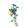
| ||||||||||||
|---|---|---|---|---|---|---|---|---|---|---|---|---|---|
| 1 |
| ||||||||||||
| Unit cell |
|
-Components
| #1: Protein | Mass: 44239.742 Da / Num. of mol.: 1 / Fragment: residues 26-405 Source method: isolated from a genetically manipulated source Source: (gene. exp.)  Homo sapiens (human) / References: UniProt: Q90922 Homo sapiens (human) / References: UniProt: Q90922 | ||||
|---|---|---|---|---|---|
| #2: Sugar |   Type: D-saccharide, beta linking / Mass: 221.208 Da / Num. of mol.: 2 Type: D-saccharide, beta linking / Mass: 221.208 Da / Num. of mol.: 2Source method: isolated from a genetically manipulated source Formula: C8H15NO6 Has ligand of interest | N | Has protein modification | Y | |
-Experimental details
-Experiment
| Experiment | Method: X-RAY DIFFRACTION / Number of used crystals: 1 |
|---|
- Sample preparation
| Crystal | Density Matthews: 4.75 Å3/Da / Density % sol: 74.09 % |
|---|---|
| Crystal grow | Temperature: 293 K / Method: vapor diffusion, sitting drop Details: 0.2 M Sodium phosphate dibasic dihydrate, 20% w/v Polyethylene glycol 3,350 |
-Data collection
| Diffraction | Mean temperature: 100 K / Serial crystal experiment: N |
|---|---|
| Diffraction source | Source: SYNCHROTRON / Site: APS  / Beamline: 23-ID-B / Wavelength: 1.0332 Å / Beamline: 23-ID-B / Wavelength: 1.0332 Å |
| Detector | Type: DECTRIS EIGER X 16M / Detector: PIXEL / Date: Mar 13, 2022 |
| Radiation | Protocol: SINGLE WAVELENGTH / Monochromatic (M) / Laue (L): M / Scattering type: x-ray |
| Radiation wavelength | Wavelength: 1.0332 Å / Relative weight: 1 |
| Reflection | Resolution: 3.9→37.77 Å / Num. obs: 13196 / % possible obs: 97.8 % / Redundancy: 13.1 % / Biso Wilson estimate: 155.68 Å2 / CC1/2: 0.999 / Rpim(I) all: 0.0705 / Net I/σ(I): 7 |
| Reflection shell | Resolution: 3.9→4.04 Å / Redundancy: 13.8 % / Mean I/σ(I) obs: 0.87 / Num. unique obs: 1682 / CC1/2: 0.705 / Rpim(I) all: 1.268 / % possible all: 96.8 |
- Processing
| Software |
| ||||||||||||||||||||||||||||||||||||||||||||||||||||||||
|---|---|---|---|---|---|---|---|---|---|---|---|---|---|---|---|---|---|---|---|---|---|---|---|---|---|---|---|---|---|---|---|---|---|---|---|---|---|---|---|---|---|---|---|---|---|---|---|---|---|---|---|---|---|---|---|---|---|
| Refinement | Method to determine structure: MOLECULAR REPLACEMENT / Resolution: 3.9→37.77 Å / SU ML: 0.5595 / Cross valid method: FREE R-VALUE / σ(F): 1.34 / Phase error: 47.8442 Stereochemistry target values: GeoStd + Monomer Library + CDL v1.2
| ||||||||||||||||||||||||||||||||||||||||||||||||||||||||
| Solvent computation | Shrinkage radii: 0.9 Å / VDW probe radii: 1.1 Å / Solvent model: FLAT BULK SOLVENT MODEL | ||||||||||||||||||||||||||||||||||||||||||||||||||||||||
| Displacement parameters | Biso mean: 216.96 Å2 | ||||||||||||||||||||||||||||||||||||||||||||||||||||||||
| Refinement step | Cycle: LAST / Resolution: 3.9→37.77 Å
| ||||||||||||||||||||||||||||||||||||||||||||||||||||||||
| Refine LS restraints |
| ||||||||||||||||||||||||||||||||||||||||||||||||||||||||
| LS refinement shell |
| ||||||||||||||||||||||||||||||||||||||||||||||||||||||||
| Refinement TLS params. | Method: refined / Origin x: 9.41483616592 Å / Origin y: -33.8144231617 Å / Origin z: -19.7101355974 Å
| ||||||||||||||||||||||||||||||||||||||||||||||||||||||||
| Refinement TLS group | Selection details: (chain 'A' and resid 40 through 405) |
