 Movie
Movie Controller


+ Open data

- Basic information
| Entry | Database: PDB / ID: 8trg | ||||||
|---|---|---|---|---|---|---|---|
| Title | Structure of full-length LexA bound to a RecA filament | ||||||
 Components Components |
| ||||||
 Keywords Keywords | SIGNALING PROTEIN/DNA / Damage response / signal transduction / SIGNALING PROTEIN / SIGNALING PROTEIN-DNA complex | ||||||
| Function / homology |  Function and homology information Function and homology informationrepressor LexA / DNA polymerase V complex / homologous recombination / SOS response / recombinational repair / DNA-binding transcription repressor activity / ATP-dependent DNA damage sensor activity / response to ionizing radiation / ATP-dependent activity, acting on DNA / translesion synthesis ...repressor LexA / DNA polymerase V complex / homologous recombination / SOS response / recombinational repair / DNA-binding transcription repressor activity / ATP-dependent DNA damage sensor activity / response to ionizing radiation / ATP-dependent activity, acting on DNA / translesion synthesis / cell motility / protein-DNA complex / single-stranded DNA binding / DNA recombination / DNA-binding transcription factor binding / sequence-specific DNA binding / damaged DNA binding / DNA replication / transcription cis-regulatory region binding / serine-type endopeptidase activity / DNA repair / negative regulation of DNA-templated transcription / DNA damage response / DNA-templated transcription / proteolysis / DNA binding / ATP binding / identical protein binding / cytoplasm / cytosol Similarity search - Function | ||||||
| Biological species |  | ||||||
| Method | ELECTRON MICROSCOPY / helical reconstruction / cryo EM / Resolution: 2.93 Å | ||||||
 Authors Authors | Cory, M.B. / Li, A. / Kohli, R.M. | ||||||
| Funding support |  United States, 1items United States, 1items
| ||||||
 Citation Citation | Journal: Nat Struct Mol Biol / Year: 2024 Title: The LexA-RecA* structure reveals a cryptic lock-and-key mechanism for SOS activation. Authors: Michael B Cory / Allen Li / Christina M Hurley / Peter J Carman / Ruth A Pumroy / Zachary M Hostetler / Ryann M Perez / Yarra Venkatesh / Xinning Li / Kushol Gupta / E James Petersson / Rahul M Kohli / Abstract: The bacterial SOS response plays a key role in adaptation to DNA damage, including genomic stress caused by antibiotics. SOS induction begins when activated RecA*, an oligomeric nucleoprotein ...The bacterial SOS response plays a key role in adaptation to DNA damage, including genomic stress caused by antibiotics. SOS induction begins when activated RecA*, an oligomeric nucleoprotein filament that forms on single-stranded DNA, binds to and stimulates autoproteolysis of the repressor LexA. Here, we present the structure of the complete Escherichia coli SOS signal complex, constituting full-length LexA bound to RecA*. We uncover an extensive interface unexpectedly including the LexA DNA-binding domain, providing a new molecular rationale for ordered SOS gene induction. We further find that the interface involves three RecA subunits, with a single residue in the central engaged subunit acting as a molecular key, inserting into an allosteric binding pocket to induce LexA cleavage. Given the pro-mutagenic nature of SOS activation, our structural and mechanistic insights provide a foundation for developing new therapeutics to slow the evolution of antibiotic resistance. | ||||||
| History |
|
- Structure visualization
| Structure viewer | Molecule: MolmilJmol/JSmol |
|---|
- Downloads & links
-Download
| PDBx/mmCIF format | 8trg.cif.gz | 1022.6 KB | Display | PDBx/mmCIF format |
|---|---|---|---|---|
| PDB format | pdb8trg.ent.gz | Display | PDB format | |
| PDBx/mmJSON format | 8trg.json.gz | Tree view | PDBx/mmJSON format | |
| Others |  Other downloads Other downloads |
-Validation report
| Arichive directory | https://data.pdbj.org/pub/pdb/validation_reports/tr/8trgftp://data.pdbj.org/pub/pdb/validation_reports/tr/8trg | HTTPS FTP |
|---|
-Related structure data
| Related structure data |  41579MC M: map data used to model this data C: citing same article ( |
|---|---|
| Similar structure data |
-Links
 PDBj
PDBj


- Assembly
| Deposited unit | 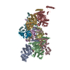
|
|---|---|
| 1 |
|
-Components
| #1: Protein | Mass: 41119.551 Da / Num. of mol.: 8 Source method: isolated from a genetically manipulated source Source: (gene. exp.) #2: Protein | Mass: 22612.887 Da / Num. of mol.: 2 / Mutation: K156A Source method: isolated from a genetically manipulated source Source: (gene. exp.) #3: DNA chain | | Mass: 8618.482 Da / Num. of mol.: 1 / Source method: obtained synthetically / Details: Synthetic oligo / Source: (synth.) #4: Chemical | ChemComp-AGS /   Mass: 523.247 Da / Num. of mol.: 8 / Source method: obtained synthetically / Formula: C10H16N5O12P3S / Comment: ATP-gamma-S, energy-carrying molecule analogue*YM Mass: 523.247 Da / Num. of mol.: 8 / Source method: obtained synthetically / Formula: C10H16N5O12P3S / Comment: ATP-gamma-S, energy-carrying molecule analogue*YM#5: Chemical | ChemComp-MG /   Mass: 24.305 Da / Num. of mol.: 8 / Source method: obtained synthetically / Formula: Mg Mass: 24.305 Da / Num. of mol.: 8 / Source method: obtained synthetically / Formula: MgHas ligand of interest | N | Has protein modification | N | |
|---|
-Experimental details
-Experiment
| Experiment | Method: ELECTRON MICROSCOPY |
|---|---|
| EM experiment | Aggregation state: FILAMENT / 3D reconstruction method: helical reconstruction |
- Sample preparation
| Component |
| ||||||||||||||||||||||||||||||
|---|---|---|---|---|---|---|---|---|---|---|---|---|---|---|---|---|---|---|---|---|---|---|---|---|---|---|---|---|---|---|---|
| Molecular weight |
| ||||||||||||||||||||||||||||||
| Source (natural) |
| ||||||||||||||||||||||||||||||
| Source (recombinant) |
| ||||||||||||||||||||||||||||||
| Buffer solution | pH: 7.5 Details: 70 mM Tris, pH 7.5, 150 mM NaCl, 1 mM MgCl2, 2 mM TCEP, 0.25 mM ATPyS | ||||||||||||||||||||||||||||||
| Buffer component |
| ||||||||||||||||||||||||||||||
| Specimen | Conc.: 0.2 mg/ml / Embedding applied: NO / Shadowing applied: NO / Staining applied: NO / Vitrification applied: YES | ||||||||||||||||||||||||||||||
| Specimen support | Details: 25 mA current / Grid material: COPPER / Grid mesh size: 300 divisions/in. / Grid type: Quantifoil R1.2/1.3 | ||||||||||||||||||||||||||||||
| Vitrification | Instrument: FEI VITROBOT MARK IV / Cryogen name: ETHANE / Humidity: 100 % / Chamber temperature: 298 K / Details: Blotting time of 7.0 s; 0.0 blot force |
- Electron microscopy imaging
| Experimental equipment |  Model: Titan Krios / Image courtesy: FEI Company |
|---|---|
| Microscopy | Model: FEI TITAN KRIOS / Details: Preliminary grid screening was done manually |
| Electron gun | Electron source:  FIELD EMISSION GUN / Accelerating voltage: 300 kV / Illumination mode: SPOT SCAN FIELD EMISSION GUN / Accelerating voltage: 300 kV / Illumination mode: SPOT SCAN |
| Electron lens | Mode: BRIGHT FIELD / Nominal magnification: 64000 X / Nominal defocus max: 2000 nm / Nominal defocus min: 500 nm / Cs: 2.7 mm / C2 aperture diameter: 100 µm |
| Specimen holder | Specimen holder model: FEI TITAN KRIOS AUTOGRID HOLDER |
| Image recording | Average exposure time: 5.35 sec. / Electron dose: 46.2 e/Å2 / Film or detector model: GATAN K3 (6k x 4k) / Num. of grids imaged: 1 / Num. of real images: 2748 / Details: 40 frames collected per movie |
- Processing
| EM software |
| |||||||||||||||||||||||||||||||||||||||||||||
|---|---|---|---|---|---|---|---|---|---|---|---|---|---|---|---|---|---|---|---|---|---|---|---|---|---|---|---|---|---|---|---|---|---|---|---|---|---|---|---|---|---|---|---|---|---|---|
| CTF correction | Type: PHASE FLIPPING AND AMPLITUDE CORRECTION | |||||||||||||||||||||||||||||||||||||||||||||
| Helical symmerty |
| |||||||||||||||||||||||||||||||||||||||||||||
| Particle selection | Num. of particles selected: 3423408 Details: cryoSPARC automated Filament Tracer using filament diameter of 100A, 17A separation between segments. Minimum and maximum filament diameters of 90A, 150A respectively. Inspect Picks to ...Details: cryoSPARC automated Filament Tracer using filament diameter of 100A, 17A separation between segments. Minimum and maximum filament diameters of 90A, 150A respectively. Inspect Picks to filter particles with high curvature or sinuosity Local power > 567.940 Local power < 5711.103 Curvature (1/A) < 0.005970 Sinuosity < 1.393924 Low pass filtered particles near micrograph edge. | |||||||||||||||||||||||||||||||||||||||||||||
| 3D reconstruction | Resolution: 2.93 Å / Resolution method: FSC 0.143 CUT-OFF / Num. of particles: 233920 / Num. of class averages: 2 / Symmetry type: HELICAL | |||||||||||||||||||||||||||||||||||||||||||||
| Atomic model building | Protocol: RIGID BODY FIT / Space: REAL | |||||||||||||||||||||||||||||||||||||||||||||
| Atomic model building | Details: The initial model was generated via benchling plugin for full-length construct sequence Source name: AlphaFold / Type: in silico model | |||||||||||||||||||||||||||||||||||||||||||||
| Refine LS restraints |
|
