 Movie
Movie Controller

[English] 日本語
 Yorodumi
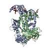
- PDB-8tp8: Structure of the C. crescentus WYL-activator, DriD, bound to ssDN... -
+ Open data

- Basic information
| Entry | Database: PDB / ID: 8tp8 | ||||||
|---|---|---|---|---|---|---|---|
| Title | Structure of the C. crescentus WYL-activator, DriD, bound to ssDNA and cognate DNA | ||||||
 Components Components |
| ||||||
 Keywords Keywords | TRANSCRIPTION/DNA / WYL motif / DriD / C. crescentus / DNA damage repair / ssDNA / transcription activation / TRANSCRIPTION / TRANSCRIPTION-DNA complex | ||||||
| Function / homology | Protein PafC / WCX domain / : / WYL domain profile. / WYL domain / WYL domain / DNA / DNA (> 10) / DeoR-family transcriptional regulator Function and homology information Function and homology information | ||||||
| Biological species |  Caulobacter vibrioides NA1000 (bacteria)Caulobacter vibrioides (bacteria) Caulobacter vibrioides NA1000 (bacteria)Caulobacter vibrioides (bacteria) | ||||||
| Method |  X-RAY DIFFRACTION / SYNCHROTRON / MOLECULAR REPLACEMENT / Resolution: 2.74 Å X-RAY DIFFRACTION / SYNCHROTRON / MOLECULAR REPLACEMENT / Resolution: 2.74 Å | ||||||
 Authors Authors | Schumacher, M.A. | ||||||
| Funding support |  United States, 1items United States, 1items
| ||||||
 Citation Citation | Journal: Nucleic Acids Res. / Year: 2024 Title: Structure of the WYL-domain containing transcription activator, DriD, in complex with ssDNA effector and DNA target site. Authors: Schumacher, M.A. / Cannistraci, E. / Salinas, R. / Lloyd, D. / Messner, E. / Gozzi, K. | ||||||
| History |
|
- Structure visualization
| Structure viewer | Molecule: MolmilJmol/JSmol |
|---|
- Downloads & links
-Download
| PDBx/mmCIF format | 8tp8.cif.gz | 615.5 KB | Display | PDBx/mmCIF format |
|---|---|---|---|---|
| PDB format | pdb8tp8.ent.gz | 502.8 KB | Display | PDB format |
| PDBx/mmJSON format | 8tp8.json.gz | Tree view | PDBx/mmJSON format | |
| Others |  Other downloads Other downloads |
-Validation report
| Arichive directory | https://data.pdbj.org/pub/pdb/validation_reports/tp/8tp8ftp://data.pdbj.org/pub/pdb/validation_reports/tp/8tp8 | HTTPS FTP |
|---|
-Related structure data
-Links
 PDBj
PDBj


- Assembly
| Deposited unit | 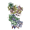
| ||||||||
|---|---|---|---|---|---|---|---|---|---|
| 1 | 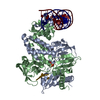
| ||||||||
| 2 | 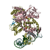
| ||||||||
| Unit cell |
|
-Components
-Protein , 1 types, 4 molecules ABCD
| #1: Protein | Mass: 38037.203 Da / Num. of mol.: 4 Source method: isolated from a genetically manipulated source Source: (gene. exp.) Caulobacter vibrioides NA1000 (bacteria)Gene: CCNA_01151 / Production host: |
|---|
-DNA chain , 3 types, 8 molecules UFRTLYJK
| #2: DNA chain | Mass: 6437.183 Da / Num. of mol.: 2 / Source method: obtained synthetically / Source: (synth.) Caulobacter vibrioides (bacteria)#3: DNA chain | Mass: 6446.197 Da / Num. of mol.: 2 / Source method: obtained synthetically / Source: (synth.) Caulobacter vibrioides (bacteria)#4: DNA chain | Mass: 877.623 Da / Num. of mol.: 4 / Source method: obtained synthetically / Source: (synth.) Caulobacter vibrioides (bacteria) |
|---|
-Non-polymers , 2 types, 114 molecules 

| #5: Chemical | ChemComp-SO4 /  Mass: 96.063 Da / Num. of mol.: 6 / Source method: obtained synthetically / Formula: SO4 Mass: 96.063 Da / Num. of mol.: 6 / Source method: obtained synthetically / Formula: SO4#6: Water | ChemComp-HOH / | Mass: 18.015 Da / Num. of mol.: 108 / Source method: isolated from a natural source / Formula: H2O |
|---|
-Details
| Has ligand of interest | N |
|---|
-Experimental details
-Experiment
| Experiment | Method: X-RAY DIFFRACTION / Number of used crystals: 1 |
|---|
- Sample preparation
| Crystal | Density Matthews: 3.25 Å3/Da / Density % sol: 62.17 % |
|---|---|
| Crystal grow | Temperature: 293 K / Method: vapor diffusion, hanging drop / Details: 0.1 M Tris, 1.0 M Citrate |
-Data collection
| Diffraction | Mean temperature: 100 K / Serial crystal experiment: N |
|---|---|
| Diffraction source | Source: SYNCHROTRON / Site: ALS / Beamline: 5.0.2 / Wavelength: 1 Å |
| Detector | Type: DECTRIS PILATUS3 6M / Detector: PIXEL / Date: Jan 3, 2022 |
| Radiation | Protocol: SINGLE WAVELENGTH / Monochromatic (M) / Laue (L): M / Scattering type: x-ray |
| Radiation wavelength | Wavelength: 1 Å / Relative weight: 1 |
| Reflection | Resolution: 2.74→69 Å / Num. obs: 56088 / % possible obs: 96.1 % / Redundancy: 3.3 % / CC1/2: 0.99 / Rmerge(I) obs: 0.092 / Rpim(I) all: 0.06 / Net I/σ(I): 7.6 |
| Reflection shell | Resolution: 2.74→2.89 Å / Rmerge(I) obs: 0.657 / Mean I/σ(I) obs: 1.5 / Num. unique obs: 3276 / CC1/2: 0.635 |
- Processing
| Software |
| |||||||||||||||||||||||||||||||||||||||||||||||||||||||||||||||||||||||||||||||||||||||||||||||||||||||||
|---|---|---|---|---|---|---|---|---|---|---|---|---|---|---|---|---|---|---|---|---|---|---|---|---|---|---|---|---|---|---|---|---|---|---|---|---|---|---|---|---|---|---|---|---|---|---|---|---|---|---|---|---|---|---|---|---|---|---|---|---|---|---|---|---|---|---|---|---|---|---|---|---|---|---|---|---|---|---|---|---|---|---|---|---|---|---|---|---|---|---|---|---|---|---|---|---|---|---|---|---|---|---|---|---|---|---|
| Refinement | Method to determine structure: MOLECULAR REPLACEMENT / Resolution: 2.74→68.97 Å / SU ML: 0.45 / Cross valid method: FREE R-VALUE / σ(F): 1.35 / Phase error: 28.59 / Stereochemistry target values: ML
| |||||||||||||||||||||||||||||||||||||||||||||||||||||||||||||||||||||||||||||||||||||||||||||||||||||||||
| Solvent computation | Shrinkage radii: 0.9 Å / VDW probe radii: 1.11 Å / Solvent model: FLAT BULK SOLVENT MODEL | |||||||||||||||||||||||||||||||||||||||||||||||||||||||||||||||||||||||||||||||||||||||||||||||||||||||||
| Refinement step | Cycle: LAST / Resolution: 2.74→68.97 Å
| |||||||||||||||||||||||||||||||||||||||||||||||||||||||||||||||||||||||||||||||||||||||||||||||||||||||||
| Refine LS restraints |
| |||||||||||||||||||||||||||||||||||||||||||||||||||||||||||||||||||||||||||||||||||||||||||||||||||||||||
| LS refinement shell |
| |||||||||||||||||||||||||||||||||||||||||||||||||||||||||||||||||||||||||||||||||||||||||||||||||||||||||
| Refinement TLS params. | Method: refined / Origin x: 29.1364 Å / Origin y: -16.2606 Å / Origin z: 45.204 Å
| |||||||||||||||||||||||||||||||||||||||||||||||||||||||||||||||||||||||||||||||||||||||||||||||||||||||||
| Refinement TLS group | Selection details: all |
