 Movie
Movie Controller


+ Open data

- Basic information
| Entry | Database: PDB / ID: 8tnv | |||||||||||||||
|---|---|---|---|---|---|---|---|---|---|---|---|---|---|---|---|---|
| Title | Hemocyanin Functional Unit CCHB-g of Concholepas concholepas | |||||||||||||||
 Components Components | Hemocyanin Functional Unit CCHB-g | |||||||||||||||
 Keywords Keywords | OXYGEN TRANSPORT / Hemocyanin / metalloprotein / copper protein / Concholepas concholepas | |||||||||||||||
| Function / homology | BROMIDE ION / CU2-O2 CLUSTER / Chem-PG6 Function and homology information Function and homology information | |||||||||||||||
| Biological species |  Concholepas concholepas (barnacle rock-shell) Concholepas concholepas (barnacle rock-shell) | |||||||||||||||
| Method |  X-RAY DIFFRACTION / SYNCHROTRON / SAD / Resolution: 1.5 Å X-RAY DIFFRACTION / SYNCHROTRON / SAD / Resolution: 1.5 Å | |||||||||||||||
 Authors Authors | Munoz, S. / Vallejos-Baccelliere, G. / Manubens, A. / Salazar, M. / Nascimento, A.F.Z. / Ambrosio, A.L.B. / Becker, M.I. / Guixe, V. / Castro-Fernandez, V. | |||||||||||||||
| Funding support |  Chile, 4items Chile, 4items
| |||||||||||||||
 Citation Citation | Journal: Structure / Year: 2024 Title: Structural insights into a functional unit from an immunogenic mollusk hemocyanin. Authors: Munoz, S.M. / Vallejos-Baccelliere, G. / Manubens, A. / Salazar, M.L. / Nascimento, A.F.Z. / Tapia-Reyes, P. / Meneses, C. / Ambrosio, A.L.B. / Becker, M.I. / Guixe, V. / Castro-Fernandez, V. | |||||||||||||||
| History |
|
- Structure visualization
| Structure viewer | Molecule: MolmilJmol/JSmol |
|---|
- Downloads & links
-Download
| PDBx/mmCIF format | 8tnv.cif.gz | 200.3 KB | Display | PDBx/mmCIF format |
|---|---|---|---|---|
| PDB format | pdb8tnv.ent.gz | 156.3 KB | Display | PDB format |
| PDBx/mmJSON format | 8tnv.json.gz | Tree view | PDBx/mmJSON format | |
| Others |  Other downloads Other downloads |
-Validation report
| Arichive directory | https://data.pdbj.org/pub/pdb/validation_reports/tn/8tnvftp://data.pdbj.org/pub/pdb/validation_reports/tn/8tnv | HTTPS FTP |
|---|
-Related structure data
| Similar structure data |
|---|
-Links
 PDBj
PDBj


- Assembly
| Deposited unit | 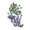
| ||||||||||
|---|---|---|---|---|---|---|---|---|---|---|---|
| 1 | 
| ||||||||||
| 2 | 
| ||||||||||
| Unit cell |
|
-Components
-Protein , 1 types, 2 molecules AB
| #1: Protein | Mass: 45285.039 Da / Num. of mol.: 2 / Source method: isolated from a natural source Source: (natural) Concholepas concholepas (barnacle rock-shell) |
|---|
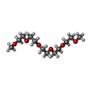
-Sugars , 2 types, 4 molecules 
| #2: Polysaccharide | 2-acetamido-2-deoxy-beta-D-glucopyranose-(1-2)-alpha-D-mannopyranose-(1-6)-beta-D-mannopyranose-(1- ...2-acetamido-2-deoxy-beta-D-glucopyranose-(1-2)-alpha-D-mannopyranose-(1-6)-beta-D-mannopyranose-(1-4)-2-acetamido-2-deoxy-beta-D-glucopyranose-(1-4)-[alpha-L-fucopyranose-(1-6)]2-acetamido-2-deoxy-beta-D-glucopyranose Source method: isolated from a genetically manipulated source |
|---|---|
| #3: Sugar |  Type: D-saccharide, beta linking / Mass: 221.208 Da / Num. of mol.: 3 / Source method: obtained synthetically / Formula: C8H15NO6 Type: D-saccharide, beta linking / Mass: 221.208 Da / Num. of mol.: 3 / Source method: obtained synthetically / Formula: C8H15NO6 |
-Non-polymers , 5 types, 933 molecules 


| #4: Chemical |  Mass: 159.091 Da / Num. of mol.: 2 / Source method: obtained synthetically / Formula: Cu2O2 Mass: 159.091 Da / Num. of mol.: 2 / Source method: obtained synthetically / Formula: Cu2O2#5: Chemical |  Mass: 79.904 Da / Num. of mol.: 3 / Source method: obtained synthetically / Formula: Br Mass: 79.904 Da / Num. of mol.: 3 / Source method: obtained synthetically / Formula: Br#6: Chemical |  Mass: 266.331 Da / Num. of mol.: 2 / Source method: obtained synthetically / Formula: C12H26O6 Mass: 266.331 Da / Num. of mol.: 2 / Source method: obtained synthetically / Formula: C12H26O6#7: Chemical | ChemComp-PG4 / |  Mass: 194.226 Da / Num. of mol.: 1 / Source method: obtained synthetically / Formula: C8H18O5 / Comment: precipitant*YM Mass: 194.226 Da / Num. of mol.: 1 / Source method: obtained synthetically / Formula: C8H18O5 / Comment: precipitant*YM#8: Water | ChemComp-HOH / | Mass: 18.015 Da / Num. of mol.: 925 / Source method: isolated from a natural source / Formula: H2O |
|---|
-Details
| Has ligand of interest | N |
|---|---|
| Has protein modification | Y |
-Experimental details
-Experiment
| Experiment | Method: X-RAY DIFFRACTION / Number of used crystals: 1 |
|---|
- Sample preparation
| Crystal | Density Matthews: 2.59 Å3/Da / Density % sol: 52.6 % |
|---|---|
| Crystal grow | Temperature: 292 K / Method: vapor diffusion, sitting drop / pH: 6 Details: Protein condition: Protein 3 mg/mL, 20 mM Imidazole-HCl pH 6.0, and 300 mM NaCl. Reservoir condition: 20% w/V PEGMME 2000 and 150 mM KBr. |
-Data collection
| Diffraction | Mean temperature: 100 K / Serial crystal experiment: N |
|---|---|
| Diffraction source | Source: SYNCHROTRON / Site: LNLS SIRIUS / Beamline: MANACA / Wavelength: 0.9772 Å |
| Detector | Type: DECTRIS PILATUS 2M / Detector: PIXEL / Date: Dec 8, 2021 |
| Radiation | Monochromator: M / Protocol: SINGLE WAVELENGTH / Monochromatic (M) / Laue (L): M / Scattering type: x-ray |
| Radiation wavelength | Wavelength: 0.9772 Å / Relative weight: 1 |
| Reflection | Resolution: 1.5→47.41 Å / Num. obs: 149523 / % possible obs: 99.92 % / Redundancy: 13.2 % / Biso Wilson estimate: 15.65 Å2 / CC1/2: 0.999 / CC star: 1 / Rmerge(I) obs: 0.1226 / Rpim(I) all: 0.03507 / Rrim(I) all: 0.1276 / Net I/σ(I): 14.47 |
| Reflection shell | Resolution: 1.5→1.554 Å / Redundancy: 13.8 % / Rmerge(I) obs: 1.411 / Mean I/σ(I) obs: 2.24 / Num. unique obs: 14807 / CC1/2: 0.762 / CC star: 0.93 / Rpim(I) all: 0.3925 / Rrim(I) all: 1.466 / % possible all: 99.99 |
- Processing
| Software |
| |||||||||||||||||||||||||||||||||||||||||||||||||||||||||||||||||||||||||||||||||||||||||||||||||||||||||||||||||||||||||||||||||||||||||||||||||||||||||||||||||||||||||||||||||||||||||||||||||||||||||||||||||||||||||
|---|---|---|---|---|---|---|---|---|---|---|---|---|---|---|---|---|---|---|---|---|---|---|---|---|---|---|---|---|---|---|---|---|---|---|---|---|---|---|---|---|---|---|---|---|---|---|---|---|---|---|---|---|---|---|---|---|---|---|---|---|---|---|---|---|---|---|---|---|---|---|---|---|---|---|---|---|---|---|---|---|---|---|---|---|---|---|---|---|---|---|---|---|---|---|---|---|---|---|---|---|---|---|---|---|---|---|---|---|---|---|---|---|---|---|---|---|---|---|---|---|---|---|---|---|---|---|---|---|---|---|---|---|---|---|---|---|---|---|---|---|---|---|---|---|---|---|---|---|---|---|---|---|---|---|---|---|---|---|---|---|---|---|---|---|---|---|---|---|---|---|---|---|---|---|---|---|---|---|---|---|---|---|---|---|---|---|---|---|---|---|---|---|---|---|---|---|---|---|---|---|---|---|---|---|---|---|---|---|---|---|---|---|---|---|---|---|---|---|
| Refinement | Method to determine structure: SAD / Resolution: 1.5→47.41 Å / SU ML: 0.152 / Cross valid method: FREE R-VALUE / σ(F): 0 / Phase error: 19.317 Stereochemistry target values: GEOSTD + MONOMER LIBRARY + CDL V1.2
| |||||||||||||||||||||||||||||||||||||||||||||||||||||||||||||||||||||||||||||||||||||||||||||||||||||||||||||||||||||||||||||||||||||||||||||||||||||||||||||||||||||||||||||||||||||||||||||||||||||||||||||||||||||||||
| Solvent computation | Shrinkage radii: 0.9 Å / VDW probe radii: 1.1 Å / Solvent model: FLAT BULK SOLVENT MODEL | |||||||||||||||||||||||||||||||||||||||||||||||||||||||||||||||||||||||||||||||||||||||||||||||||||||||||||||||||||||||||||||||||||||||||||||||||||||||||||||||||||||||||||||||||||||||||||||||||||||||||||||||||||||||||
| Displacement parameters | Biso mean: 21.3 Å2 | |||||||||||||||||||||||||||||||||||||||||||||||||||||||||||||||||||||||||||||||||||||||||||||||||||||||||||||||||||||||||||||||||||||||||||||||||||||||||||||||||||||||||||||||||||||||||||||||||||||||||||||||||||||||||
| Refinement step | Cycle: LAST / Resolution: 1.5→47.41 Å
| |||||||||||||||||||||||||||||||||||||||||||||||||||||||||||||||||||||||||||||||||||||||||||||||||||||||||||||||||||||||||||||||||||||||||||||||||||||||||||||||||||||||||||||||||||||||||||||||||||||||||||||||||||||||||
| Refine LS restraints |
| |||||||||||||||||||||||||||||||||||||||||||||||||||||||||||||||||||||||||||||||||||||||||||||||||||||||||||||||||||||||||||||||||||||||||||||||||||||||||||||||||||||||||||||||||||||||||||||||||||||||||||||||||||||||||
| LS refinement shell |
|
