 Movie
Movie Controller

[English] 日本語
 Yorodumi
- PDB-8tn8: Crystal structure of the murine astrovirus capsid spike at 1.75 A -
+ Open data

- Basic information
| Entry | Database: PDB / ID: 8tn8 | ||||||
|---|---|---|---|---|---|---|---|
| Title | Crystal structure of the murine astrovirus capsid spike at 1.75 A | ||||||
 Components Components | Capsid polyprotein VP90 | ||||||
 Keywords Keywords | VIRAL PROTEIN / capsid / spike / attachment / homodimer | ||||||
| Function / homology | Capsid, astroviral / Astrovirus capsid protein nucleoplasmin-like domain / Viral coat protein subunit / : / Capsid polyprotein VP90 Function and homology information Function and homology information | ||||||
| Biological species |  Murine astrovirus Murine astrovirus | ||||||
| Method |  X-RAY DIFFRACTION / SYNCHROTRON / MOLECULAR REPLACEMENT / Resolution: 1.75 Å X-RAY DIFFRACTION / SYNCHROTRON / MOLECULAR REPLACEMENT / Resolution: 1.75 Å | ||||||
 Authors Authors | Lanning, S. / DuBois, R.M. | ||||||
| Funding support |  United States, 1items United States, 1items
| ||||||
 Citation Citation | Journal: J.Gen.Virol. / Year: 2023 Title: Structure and immunogenicity of the murine astrovirus capsid spike. Authors: Lanning, S. / Pedicino, N. / Haley, D.J. / Hernandez, S. / Cortez, V. / DuBois, R.M. | ||||||
| History |
|
- Structure visualization
| Structure viewer | Molecule: MolmilJmol/JSmol |
|---|
- Downloads & links
-Download
| PDBx/mmCIF format | 8tn8.cif.gz | 249.2 KB | Display | PDBx/mmCIF format |
|---|---|---|---|---|
| PDB format | pdb8tn8.ent.gz | 178.1 KB | Display | PDB format |
| PDBx/mmJSON format | 8tn8.json.gz | Tree view | PDBx/mmJSON format | |
| Others |  Other downloads Other downloads |
-Validation report
| Arichive directory | https://data.pdbj.org/pub/pdb/validation_reports/tn/8tn8ftp://data.pdbj.org/pub/pdb/validation_reports/tn/8tn8 | HTTPS FTP |
|---|
-Related structure data
| Similar structure data |
|---|
-Links
 PDBj
PDBj- Assembly
| Deposited unit | 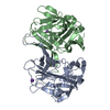
| ||||||||||||
|---|---|---|---|---|---|---|---|---|---|---|---|---|---|
| 1 |
| ||||||||||||
| Unit cell |
|
-Components
| #1: Protein | Mass: 29257.432 Da / Num. of mol.: 2 Source method: isolated from a genetically manipulated source Source: (gene. exp.) Murine astrovirus / Production host:  #2: Chemical |   Mass: 39.098 Da / Num. of mol.: 2 / Source method: obtained synthetically / Formula: K Mass: 39.098 Da / Num. of mol.: 2 / Source method: obtained synthetically / Formula: K#3: Water | ChemComp-HOH / |  Mass: 18.015 Da / Num. of mol.: 322 / Source method: isolated from a natural source / Formula: H2O Mass: 18.015 Da / Num. of mol.: 322 / Source method: isolated from a natural source / Formula: H2OHas ligand of interest | N | |
|---|
-Experimental details
-Experiment
| Experiment | Method: X-RAY DIFFRACTION / Number of used crystals: 1 |
|---|
- Sample preparation
| Crystal | Density Matthews: 2.16 Å3/Da / Density % sol: 43.11 % / Description: Small rod shaped crystal |
|---|---|
| Crystal grow | Temperature: 295.15 K / Method: vapor diffusion, sitting drop / Details: Potassium formate, PEG 3350 / PH range: 7.3-8 |
-Data collection
| Diffraction | Mean temperature: 100 K / Serial crystal experiment: N |
|---|---|
| Diffraction source | Source: SYNCHROTRON / Site: APS / Beamline: 23-ID-D / Wavelength: 1.0332 Å |
| Detector | Type: DECTRIS PILATUS3 6M / Detector: PIXEL / Date: Dec 10, 2021 |
| Radiation | Protocol: SINGLE WAVELENGTH / Monochromatic (M) / Laue (L): M / Scattering type: x-ray |
| Radiation wavelength | Wavelength: 1.0332 Å / Relative weight: 1 |
| Reflection | Resolution: 1.75→43.76 Å / Num. obs: 46845 / % possible obs: 94.79 % / Redundancy: 1.7 % / Biso Wilson estimate: 19.93 Å2 / CC1/2: 0.995 / Net I/σ(I): 14.4 |
| Reflection shell | Resolution: 1.75→1.78 Å / Redundancy: 1.7 % / Mean I/σ(I) obs: 1.9 / Num. unique obs: 2226 / CC1/2: 0.936 / % possible all: 92.52 |
- Processing
| Software |
| |||||||||||||||||||||||||||||||||||||||||||||||||||||||||||||||||||||||||||||||||||||||||||||||||||||||||||||||||||||||
|---|---|---|---|---|---|---|---|---|---|---|---|---|---|---|---|---|---|---|---|---|---|---|---|---|---|---|---|---|---|---|---|---|---|---|---|---|---|---|---|---|---|---|---|---|---|---|---|---|---|---|---|---|---|---|---|---|---|---|---|---|---|---|---|---|---|---|---|---|---|---|---|---|---|---|---|---|---|---|---|---|---|---|---|---|---|---|---|---|---|---|---|---|---|---|---|---|---|---|---|---|---|---|---|---|---|---|---|---|---|---|---|---|---|---|---|---|---|---|---|---|
| Refinement | Method to determine structure: MOLECULAR REPLACEMENT / Resolution: 1.75→43.75 Å / SU ML: 0.1755 / Cross valid method: FREE R-VALUE / σ(F): 2 / Phase error: 21.8905 Stereochemistry target values: GeoStd + Monomer Library + CDL v1.2
| |||||||||||||||||||||||||||||||||||||||||||||||||||||||||||||||||||||||||||||||||||||||||||||||||||||||||||||||||||||||
| Solvent computation | Shrinkage radii: 0.9 Å / VDW probe radii: 1.1 Å / Solvent model: FLAT BULK SOLVENT MODEL | |||||||||||||||||||||||||||||||||||||||||||||||||||||||||||||||||||||||||||||||||||||||||||||||||||||||||||||||||||||||
| Displacement parameters | Biso mean: 27.11 Å2 | |||||||||||||||||||||||||||||||||||||||||||||||||||||||||||||||||||||||||||||||||||||||||||||||||||||||||||||||||||||||
| Refinement step | Cycle: LAST / Resolution: 1.75→43.75 Å
| |||||||||||||||||||||||||||||||||||||||||||||||||||||||||||||||||||||||||||||||||||||||||||||||||||||||||||||||||||||||
| Refine LS restraints |
| |||||||||||||||||||||||||||||||||||||||||||||||||||||||||||||||||||||||||||||||||||||||||||||||||||||||||||||||||||||||
| LS refinement shell |
|
