 Movie
Movie Controller

[English] 日本語
 Yorodumi
- PDB-8tms: Crystal structure of bacterial pectin methylesterase PmeC2 from r... -
+ Open data

- Basic information
| Entry | Database: PDB / ID: 8tms | ||||||
|---|---|---|---|---|---|---|---|
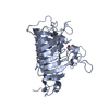
| Title | Crystal structure of bacterial pectin methylesterase PmeC2 from rumen Butyrivibrio | ||||||
 Components Components | Pectinesterase | ||||||
 Keywords Keywords | SUGAR BINDING PROTEIN / Pectin methylesterase / Butyrivibrio / rumen / pectin / methanol / methane | ||||||
| Function / homology |  Function and homology information Function and homology informationpectinesterase / pectinesterase activity / cell wall modification / pectin catabolic process / cell outer membrane Similarity search - Function | ||||||
| Biological species |  Butyrivibrio fibrisolvens (bacteria) Butyrivibrio fibrisolvens (bacteria) | ||||||
| Method |  X-RAY DIFFRACTION / SYNCHROTRON / MOLECULAR REPLACEMENT / Resolution: 2.3 Å X-RAY DIFFRACTION / SYNCHROTRON / MOLECULAR REPLACEMENT / Resolution: 2.3 Å | ||||||
 Authors Authors | Carbone, V. / Reilly, K. / Sang, C. / Schofield, L. / Ronimus, R. / Kelly, W.J. / Attwood, G.T. / Palevich, N. | ||||||
| Funding support |  New Zealand, 1items New Zealand, 1items
| ||||||
 Citation Citation | Journal: Int J Mol Sci / Year: 2023 Title: Crystal Structures of Bacterial Pectin Methylesterases Pme8A and PmeC2 from Rumen Butyrivibrio . Authors: Carbone, V. / Reilly, K. / Sang, C. / Schofield, L.R. / Ronimus, R.S. / Kelly, W.J. / Attwood, G.T. / Palevich, N. | ||||||
| History |
|
- Structure visualization
| Structure viewer | Molecule: MolmilJmol/JSmol |
|---|
- Downloads & links
-Download
| PDBx/mmCIF format | 8tms.cif.gz | 475.2 KB | Display | PDBx/mmCIF format |
|---|---|---|---|---|
| PDB format | pdb8tms.ent.gz | 391.2 KB | Display | PDB format |
| PDBx/mmJSON format | 8tms.json.gz | Tree view | PDBx/mmJSON format | |
| Others |  Other downloads Other downloads |
-Validation report
| Arichive directory | https://data.pdbj.org/pub/pdb/validation_reports/tm/8tmsftp://data.pdbj.org/pub/pdb/validation_reports/tm/8tms | HTTPS FTP |
|---|
-Related structure data
-Links
 PDBj
PDBj- Assembly
| Deposited unit | 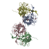
| ||||||||
|---|---|---|---|---|---|---|---|---|---|
| 1 | 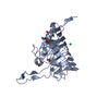
| ||||||||
| 2 | 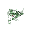
| ||||||||
| 3 | 
| ||||||||
| 4 | 
| ||||||||
| Unit cell |
|
-Components
| #1: Protein | Mass: 34418.730 Da / Num. of mol.: 4 Source method: isolated from a genetically manipulated source Source: (gene. exp.) Butyrivibrio fibrisolvens (bacteria) / Gene: SAMN02745229_01989 / Production host: #2: Chemical |   Mass: 35.453 Da / Num. of mol.: 2 / Source method: obtained synthetically / Formula: Cl Mass: 35.453 Da / Num. of mol.: 2 / Source method: obtained synthetically / Formula: Cl#3: Chemical | ChemComp-EDO /   Mass: 62.068 Da / Num. of mol.: 17 / Source method: isolated from a natural source / Formula: C2H6O2 Mass: 62.068 Da / Num. of mol.: 17 / Source method: isolated from a natural source / Formula: C2H6O2#4: Water | ChemComp-HOH / |  Mass: 18.015 Da / Num. of mol.: 334 / Source method: isolated from a natural source / Formula: H2O Mass: 18.015 Da / Num. of mol.: 334 / Source method: isolated from a natural source / Formula: H2OHas ligand of interest | N | |
|---|
-Experimental details
-Experiment
| Experiment | Method: X-RAY DIFFRACTION / Number of used crystals: 1 |
|---|
- Sample preparation
| Crystal | Density Matthews: 2.57 Å3/Da / Density % sol: 51.7 % |
|---|---|
| Crystal grow | Temperature: 298 K / Method: vapor diffusion, sitting drop / pH: 8.5 Details: 0.2 M Magnesium chloride hexahydrate, 0.1 M Tris pH 8.5, 20 % w/v PEG 8000 |
-Data collection
| Diffraction | Mean temperature: 100 K / Serial crystal experiment: N |
|---|---|
| Diffraction source | Source: SYNCHROTRON / Site: Australian Synchrotron  / Beamline: MX1 / Wavelength: 0.95372 Å / Beamline: MX1 / Wavelength: 0.95372 Å |
| Detector | Type: DECTRIS EIGER2 S 9M / Detector: PIXEL / Date: Mar 31, 2023 |
| Radiation | Protocol: SINGLE WAVELENGTH / Monochromatic (M) / Laue (L): M / Scattering type: x-ray |
| Radiation wavelength | Wavelength: 0.95372 Å / Relative weight: 1 |
| Reflection | Resolution: 2.3→47.2 Å / Num. obs: 56000 / % possible obs: 95.1 % / Redundancy: 3.7 % / CC1/2: 0.994 / Rmerge(I) obs: 0.148 / Rpim(I) all: 0.089 / Rrim(I) all: 0.173 / Χ2: 0.49 / Net I/σ(I): 5.4 |
| Reflection shell | Resolution: 2.3→2.37 Å / % possible obs: 92.3 % / Redundancy: 3.6 % / Rmerge(I) obs: 1.404 / Num. measured all: 16208 / Num. unique obs: 4465 / CC1/2: 0.349 / Rpim(I) all: 0.842 / Rrim(I) all: 1.639 / Χ2: 0.47 / Net I/σ(I) obs: 0.6 |
- Processing
| Software |
| ||||||||||||||||||||||||||||||||||||||||||||||||||||||||||||||||||||||||||||||||||||||||||||||||||||||||||||||||||||||||||||||||||||||||||||||||||||||||||||||||||||||||||||||||||||||
|---|---|---|---|---|---|---|---|---|---|---|---|---|---|---|---|---|---|---|---|---|---|---|---|---|---|---|---|---|---|---|---|---|---|---|---|---|---|---|---|---|---|---|---|---|---|---|---|---|---|---|---|---|---|---|---|---|---|---|---|---|---|---|---|---|---|---|---|---|---|---|---|---|---|---|---|---|---|---|---|---|---|---|---|---|---|---|---|---|---|---|---|---|---|---|---|---|---|---|---|---|---|---|---|---|---|---|---|---|---|---|---|---|---|---|---|---|---|---|---|---|---|---|---|---|---|---|---|---|---|---|---|---|---|---|---|---|---|---|---|---|---|---|---|---|---|---|---|---|---|---|---|---|---|---|---|---|---|---|---|---|---|---|---|---|---|---|---|---|---|---|---|---|---|---|---|---|---|---|---|---|---|---|---|
| Refinement | Method to determine structure: MOLECULAR REPLACEMENT / Resolution: 2.3→47.17 Å / Cor.coef. Fo:Fc: 0.949 / Cor.coef. Fo:Fc free: 0.928 / SU B: 24.799 / SU ML: 0.26 / Cross valid method: THROUGHOUT / ESU R: 0.391 / ESU R Free: 0.243 / Stereochemistry target values: MAXIMUM LIKELIHOOD / Details: HYDROGENS HAVE BEEN ADDED IN THE RIDING POSITIONS
| ||||||||||||||||||||||||||||||||||||||||||||||||||||||||||||||||||||||||||||||||||||||||||||||||||||||||||||||||||||||||||||||||||||||||||||||||||||||||||||||||||||||||||||||||||||||
| Solvent computation | Ion probe radii: 0.8 Å / Shrinkage radii: 0.8 Å / VDW probe radii: 1.2 Å / Solvent model: MASK | ||||||||||||||||||||||||||||||||||||||||||||||||||||||||||||||||||||||||||||||||||||||||||||||||||||||||||||||||||||||||||||||||||||||||||||||||||||||||||||||||||||||||||||||||||||||
| Displacement parameters | Biso mean: 42.514 Å2
| ||||||||||||||||||||||||||||||||||||||||||||||||||||||||||||||||||||||||||||||||||||||||||||||||||||||||||||||||||||||||||||||||||||||||||||||||||||||||||||||||||||||||||||||||||||||
| Refinement step | Cycle: 1 / Resolution: 2.3→47.17 Å
| ||||||||||||||||||||||||||||||||||||||||||||||||||||||||||||||||||||||||||||||||||||||||||||||||||||||||||||||||||||||||||||||||||||||||||||||||||||||||||||||||||||||||||||||||||||||
| Refine LS restraints |
|