 Movie
Movie Controller


+ Open data

- Basic information
| Entry | Database: PDB / ID: 8tl1 | ||||||
|---|---|---|---|---|---|---|---|
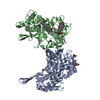
| Title | Structure of Orthoreovirus RNA Chaperone SigmaNS N17 | ||||||
 Components Components | Protein sigma-NS | ||||||
 Keywords Keywords | RNA BINDING PROTEIN / RNA-binding protein / RNA chaperone | ||||||
| Function / homology | Reovirus non-structural protein sigma NS / Sigma NS protein / host cell cytoplasm / single-stranded RNA binding / RNA-directed RNA polymerase activity / GLYCOCHOLIC ACID / Protein sigma-NS Function and homology information Function and homology information | ||||||
| Biological species |  Orthoreovirus Orthoreovirus | ||||||
| Method |  X-RAY DIFFRACTION / SYNCHROTRON / MOLECULAR REPLACEMENT / Resolution: 3.16 Å X-RAY DIFFRACTION / SYNCHROTRON / MOLECULAR REPLACEMENT / Resolution: 3.16 Å | ||||||
 Authors Authors | Prasad, B.V.V. / Zhao, B. / Hu, L. | ||||||
| Funding support |  United States, 1items United States, 1items
| ||||||
 Citation Citation | Journal: Nat Commun / Year: 2024 Title: Structure of orthoreovirus RNA chaperone sigma NS, a component of viral replication factories. Authors: Zhao, B. / Hu, L. / Kaundal, S. / Neetu, N. / Lee, C.H. / Somoulay, X. / Sankaran, B. / Taylor, G.M. / Dermody, T.S. / Venkataram Prasad, B.V. | ||||||
| History |
|
- Structure visualization
| Structure viewer | Molecule: MolmilJmol/JSmol |
|---|
- Downloads & links
-Download
| PDBx/mmCIF format | 8tl1.cif.gz | 83.3 KB | Display | PDBx/mmCIF format |
|---|---|---|---|---|
| PDB format | pdb8tl1.ent.gz | 60.2 KB | Display | PDB format |
| PDBx/mmJSON format | 8tl1.json.gz | Tree view | PDBx/mmJSON format | |
| Others |  Other downloads Other downloads |
-Validation report
| Arichive directory | https://data.pdbj.org/pub/pdb/validation_reports/tl/8tl1ftp://data.pdbj.org/pub/pdb/validation_reports/tl/8tl1 | HTTPS FTP |
|---|
-Related structure data

-Links
 PDBj
PDBj- Assembly
| Deposited unit | 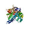
| ||||||||
|---|---|---|---|---|---|---|---|---|---|
| 1 |
| ||||||||
| Unit cell |
|
-Components
| #1: Protein | Mass: 39257.469 Da / Num. of mol.: 1 Source method: isolated from a genetically manipulated source Source: (gene. exp.) Orthoreovirus / Gene: S3 / Production host:  | ||||
|---|---|---|---|---|---|
| #2: Chemical |   Mass: 465.623 Da / Num. of mol.: 2 / Source method: obtained synthetically / Formula: C26H43NO6 / Feature type: SUBJECT OF INVESTIGATION Mass: 465.623 Da / Num. of mol.: 2 / Source method: obtained synthetically / Formula: C26H43NO6 / Feature type: SUBJECT OF INVESTIGATION#3: Water | ChemComp-HOH / |  Mass: 18.015 Da / Num. of mol.: 5 / Source method: isolated from a natural source / Formula: H2O Mass: 18.015 Da / Num. of mol.: 5 / Source method: isolated from a natural source / Formula: H2OHas ligand of interest | Y | |
-Experimental details
-Experiment
| Experiment | Method: X-RAY DIFFRACTION / Number of used crystals: 1 |
|---|
- Sample preparation
| Crystal | Density Matthews: 3.37 Å3/Da / Density % sol: 63.49 % |
|---|---|
| Crystal grow | Temperature: 293 K / Method: vapor diffusion, hanging drop / pH: 8.5 Details: 1.2% colic acid derivativemix, 0.1 M buffer system 3 (8.5), 30% precipitant Mix3 (Molecular dimensions) |
-Data collection
| Diffraction | Mean temperature: 273 K / Serial crystal experiment: N |
|---|---|
| Diffraction source | Source: SYNCHROTRON / Site: ALS / Beamline: 5.0.1 / Wavelength: 1 Å |
| Detector | Type: DECTRIS PILATUS3 2M / Detector: PIXEL / Date: May 5, 2021 |
| Radiation | Protocol: SINGLE WAVELENGTH / Monochromatic (M) / Laue (L): M / Scattering type: x-ray |
| Radiation wavelength | Wavelength: 1 Å / Relative weight: 1 |
| Reflection | Resolution: 3.16→88.84 Å / Num. obs: 7841 / % possible obs: 86.39 % / Redundancy: 1 % / Rmerge(I) obs: 0.1319 / Net I/σ(I): 10.09 |
| Reflection shell | Resolution: 3.2→3.2 Å / Rmerge(I) obs: 0.13 / Num. unique obs: 302 / Rpim(I) all: 0.13 / Rsym value: 0.18 |
- Processing
| Software |
| ||||||||||||||||||||||||||||||||||||||||||||||||||||||||||||||||||||||||||||||||||||||||||||||||||||||||||||||||||||||||||||||||||||||||||||||||||||||||||||||||||||||||||||||||||||||
|---|---|---|---|---|---|---|---|---|---|---|---|---|---|---|---|---|---|---|---|---|---|---|---|---|---|---|---|---|---|---|---|---|---|---|---|---|---|---|---|---|---|---|---|---|---|---|---|---|---|---|---|---|---|---|---|---|---|---|---|---|---|---|---|---|---|---|---|---|---|---|---|---|---|---|---|---|---|---|---|---|---|---|---|---|---|---|---|---|---|---|---|---|---|---|---|---|---|---|---|---|---|---|---|---|---|---|---|---|---|---|---|---|---|---|---|---|---|---|---|---|---|---|---|---|---|---|---|---|---|---|---|---|---|---|---|---|---|---|---|---|---|---|---|---|---|---|---|---|---|---|---|---|---|---|---|---|---|---|---|---|---|---|---|---|---|---|---|---|---|---|---|---|---|---|---|---|---|---|---|---|---|---|---|
| Refinement | Method to determine structure: MOLECULAR REPLACEMENT / Resolution: 3.16→88.84 Å / Cor.coef. Fo:Fc: 0.921 / Cor.coef. Fo:Fc free: 0.89 / SU B: 24.65 / SU ML: 0.406 / Cross valid method: THROUGHOUT / ESU R Free: 0.514 / Stereochemistry target values: MAXIMUM LIKELIHOOD / Details: HYDROGENS HAVE BEEN ADDED IN THE RIDING POSITIONS
| ||||||||||||||||||||||||||||||||||||||||||||||||||||||||||||||||||||||||||||||||||||||||||||||||||||||||||||||||||||||||||||||||||||||||||||||||||||||||||||||||||||||||||||||||||||||
| Solvent computation | Ion probe radii: 0.8 Å / Shrinkage radii: 0.8 Å / VDW probe radii: 1.2 Å / Solvent model: MASK | ||||||||||||||||||||||||||||||||||||||||||||||||||||||||||||||||||||||||||||||||||||||||||||||||||||||||||||||||||||||||||||||||||||||||||||||||||||||||||||||||||||||||||||||||||||||
| Displacement parameters | Biso mean: 65.755 Å2
| ||||||||||||||||||||||||||||||||||||||||||||||||||||||||||||||||||||||||||||||||||||||||||||||||||||||||||||||||||||||||||||||||||||||||||||||||||||||||||||||||||||||||||||||||||||||
| Refinement step | Cycle: 1 / Resolution: 3.16→88.84 Å
| ||||||||||||||||||||||||||||||||||||||||||||||||||||||||||||||||||||||||||||||||||||||||||||||||||||||||||||||||||||||||||||||||||||||||||||||||||||||||||||||||||||||||||||||||||||||
| Refine LS restraints |
|