 Movie
Movie Controller

[English] 日本語
 Yorodumi
- PDB-8tfy: tRNA 2'-phosphotransferase (Tpt1) from Pyrococcus horikoshii in c... -
+ Open data

- Basic information
| Entry | Database: PDB / ID: 8tfy | |||||||||||||||||||||
|---|---|---|---|---|---|---|---|---|---|---|---|---|---|---|---|---|---|---|---|---|---|---|
| Title | tRNA 2'-phosphotransferase (Tpt1) from Pyrococcus horikoshii in complex with NADP | |||||||||||||||||||||
 Components Components | Probable RNA 2'-phosphotransferase | |||||||||||||||||||||
 Keywords Keywords | TRANSFERASE / tRNA 2'-PHOSPHOTRANSFERASE / TPT1 / tRNA SPLICING / NADP / NAD | |||||||||||||||||||||
| Function / homology |  Function and homology information Function and homology informationtRNA 2'-phosphotransferase activity / Transferases; Transferring phosphorus-containing groups; Phosphotransferases with an alcohol group as acceptor / tRNA splicing, via endonucleolytic cleavage and ligation / NAD+ poly-ADP-ribosyltransferase activity Similarity search - Function | |||||||||||||||||||||
| Biological species |   Pyrococcus horikoshii OT3 (archaea) Pyrococcus horikoshii OT3 (archaea) | |||||||||||||||||||||
| Method |  X-RAY DIFFRACTION / SYNCHROTRON / MOLECULAR REPLACEMENT / Resolution: 1.54 Å X-RAY DIFFRACTION / SYNCHROTRON / MOLECULAR REPLACEMENT / Resolution: 1.54 Å | |||||||||||||||||||||
 Authors Authors | Jacewicz, A. / Dantuluri, S. / Shuman, S. | |||||||||||||||||||||
| Funding support |  United States, 6items United States, 6items
| |||||||||||||||||||||
 Citation Citation | Journal: Proc.Natl.Acad.Sci.USA / Year: 2023 Title: Structural basis for Tpt1-catalyzed 2'-PO 4 transfer from RNA and NADP(H) to NAD. Authors: Jacewicz, A. / Dantuluri, S. / Shuman, S. | |||||||||||||||||||||
| History |
|
- Structure visualization
| Structure viewer | Molecule: MolmilJmol/JSmol |
|---|
- Downloads & links
-Download
| PDBx/mmCIF format | 8tfy.cif.gz | 89.9 KB | Display | PDBx/mmCIF format |
|---|---|---|---|---|
| PDB format | pdb8tfy.ent.gz | 65.8 KB | Display | PDB format |
| PDBx/mmJSON format | 8tfy.json.gz | Tree view | PDBx/mmJSON format | |
| Others |  Other downloads Other downloads |
-Validation report
| Arichive directory | https://data.pdbj.org/pub/pdb/validation_reports/tf/8tfyftp://data.pdbj.org/pub/pdb/validation_reports/tf/8tfy | HTTPS FTP |
|---|
-Related structure data
| Related structure data | 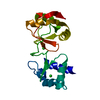 8tfiSC 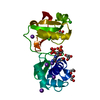 8tfxC 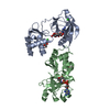 8tfzC 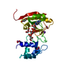 8tg3C 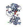 8tg4C  8tg5C 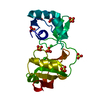 8tg6C S: Starting model for refinement C: citing same article ( |
|---|---|
| Similar structure data |
-Links
 PDBj
PDBj- Assembly
| Deposited unit | 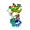
| ||||||||||
|---|---|---|---|---|---|---|---|---|---|---|---|
| 1 |
| ||||||||||
| Unit cell |
|
-Components
| #1: Protein | Mass: 20839.215 Da / Num. of mol.: 1 Source method: isolated from a genetically manipulated source Source: (gene. exp.) Pyrococcus horikoshii OT3 (archaea) / Gene: kptA, PH0160 / Plasmid: pET28b-His10Smt3 / Production host:  References: UniProt: O57899, Transferases; Transferring phosphorus-containing groups; Phosphotransferases with an alcohol group as acceptor | ||
|---|---|---|---|
| #2: Chemical | ChemComp-NAP /   Mass: 743.405 Da / Num. of mol.: 1 / Source method: obtained synthetically / Formula: C21H28N7O17P3 Mass: 743.405 Da / Num. of mol.: 1 / Source method: obtained synthetically / Formula: C21H28N7O17P3 | ||
| #3: Chemical |   Mass: 35.453 Da / Num. of mol.: 2 / Source method: obtained synthetically / Formula: Cl Mass: 35.453 Da / Num. of mol.: 2 / Source method: obtained synthetically / Formula: Cl#4: Water | ChemComp-HOH / |  Mass: 18.015 Da / Num. of mol.: 103 / Source method: isolated from a natural source / Formula: H2O Mass: 18.015 Da / Num. of mol.: 103 / Source method: isolated from a natural source / Formula: H2O |
-Experimental details
-Experiment
| Experiment | Method: X-RAY DIFFRACTION / Number of used crystals: 1 |
|---|
- Sample preparation
| Crystal | Density Matthews: 2.58 Å3/Da / Density % sol: 52.3 % |
|---|---|
| Crystal grow | Temperature: 295 K / Method: vapor diffusion, sitting drop Details: 0.1-0.3 M Tris-Bicine (pH 8.3 or 8.5), 0.1 M amino acid additives mixture (0.02 M each of DL-glutamic acid monohydrate, DL-alanine, glycine, DL-lysine monohydrate and DL-serine), 16-29.6% ...Details: 0.1-0.3 M Tris-Bicine (pH 8.3 or 8.5), 0.1 M amino acid additives mixture (0.02 M each of DL-glutamic acid monohydrate, DL-alanine, glycine, DL-lysine monohydrate and DL-serine), 16-29.6% ethylene glycol, and 8-14.8% PEG-8000 |
-Data collection
| Diffraction | Mean temperature: 100 K / Serial crystal experiment: N |
|---|---|
| Diffraction source | Source: SYNCHROTRON / Site: NSLS-II / Beamline: 19-ID / Wavelength: 0.9795 Å |
| Detector | Type: DECTRIS EIGER2 XE 9M / Detector: PIXEL / Date: Jun 16, 2023 |
| Radiation | Protocol: SINGLE WAVELENGTH / Monochromatic (M) / Laue (L): M / Scattering type: x-ray |
| Radiation wavelength | Wavelength: 0.9795 Å / Relative weight: 1 |
| Reflection | Resolution: 1.54→50 Å / Num. obs: 33140 / % possible obs: 100 % / Redundancy: 9.3 % / Biso Wilson estimate: 20.18 Å2 / CC1/2: 0.997 / Rmerge(I) obs: 0.073 / Rpim(I) all: 0.024 / Net I/σ(I): 42.5 |
| Reflection shell | Resolution: 1.54→1.57 Å / Redundancy: 6.4 % / Rmerge(I) obs: 1.005 / Num. unique obs: 1611 / CC1/2: 0.807 / Rpim(I) all: 0.398 / % possible all: 99.9 |
- Processing
| Software |
| ||||||||||||||||||||||||||||||||||||||||||||||||||||||||||||||||||||||||||||||||||||
|---|---|---|---|---|---|---|---|---|---|---|---|---|---|---|---|---|---|---|---|---|---|---|---|---|---|---|---|---|---|---|---|---|---|---|---|---|---|---|---|---|---|---|---|---|---|---|---|---|---|---|---|---|---|---|---|---|---|---|---|---|---|---|---|---|---|---|---|---|---|---|---|---|---|---|---|---|---|---|---|---|---|---|---|---|---|
| Refinement | Method to determine structure: MOLECULAR REPLACEMENT Starting model: 8TFI Resolution: 1.54→29.2 Å / SU ML: 0.142 / Cross valid method: FREE R-VALUE / σ(F): 1.36 / Phase error: 19.271 Stereochemistry target values: GEOSTD + MONOMER LIBRARY + CDL V1.2
| ||||||||||||||||||||||||||||||||||||||||||||||||||||||||||||||||||||||||||||||||||||
| Solvent computation | Shrinkage radii: 0.9 Å / VDW probe radii: 1.1 Å / Solvent model: FLAT BULK SOLVENT MODEL | ||||||||||||||||||||||||||||||||||||||||||||||||||||||||||||||||||||||||||||||||||||
| Displacement parameters | Biso mean: 27.53 Å2 | ||||||||||||||||||||||||||||||||||||||||||||||||||||||||||||||||||||||||||||||||||||
| Refinement step | Cycle: LAST / Resolution: 1.54→29.2 Å
| ||||||||||||||||||||||||||||||||||||||||||||||||||||||||||||||||||||||||||||||||||||
| Refine LS restraints |
| ||||||||||||||||||||||||||||||||||||||||||||||||||||||||||||||||||||||||||||||||||||
| LS refinement shell |
|
