 Movie
Movie Controller

[English] 日本語
 Yorodumi
- PDB-8tfs: Crystal structure of the Rhodanese-like domain of Vretifemale_108... -
+ Open data

- Basic information
| Entry | Database: PDB / ID: 8tfs | ||||||
|---|---|---|---|---|---|---|---|
| Title | Crystal structure of the Rhodanese-like domain of Vretifemale_1082 from Volvox reticuliferus | ||||||
 Components Components | Rhodanese domain-containing protein | ||||||
 Keywords Keywords | TRANSFERASE / Rhodanese / sulfur transferase | ||||||
| Function / homology | Calcium sensing receptor, plant / de-etiolation / regulation of stomatal closure / Rhodanese-like domain / Rhodanese domain profile. / Rhodanese-like domain superfamily / Rhodanese-like domain / cellular response to calcium ion / Rhodanese domain-containing protein Function and homology information Function and homology information | ||||||
| Biological species |  Volvox reticuliferus (plant) Volvox reticuliferus (plant) | ||||||
| Method |  X-RAY DIFFRACTION / SYNCHROTRON / MOLECULAR REPLACEMENT / Resolution: 1.95 Å X-RAY DIFFRACTION / SYNCHROTRON / MOLECULAR REPLACEMENT / Resolution: 1.95 Å | ||||||
 Authors Authors | Arbing, M.A. / Jonikas, M. / Yeates, T.O. | ||||||
| Funding support |  United States, 1items United States, 1items
| ||||||
 Citation Citation | Journal: To Be Published Title: Crystal structure of the Rhodanese-like domain of Vretifemale_1082 from Volvox reticuliferus Authors: Arbing, M.A. / Jonikas, M. / Yeates, T.O. | ||||||
| History |
|
- Structure visualization
| Structure viewer | Molecule: MolmilJmol/JSmol |
|---|
- Downloads & links
-Download
| PDBx/mmCIF format | 8tfs.cif.gz | 206.5 KB | Display | PDBx/mmCIF format |
|---|---|---|---|---|
| PDB format | pdb8tfs.ent.gz | 138.7 KB | Display | PDB format |
| PDBx/mmJSON format | 8tfs.json.gz | Tree view | PDBx/mmJSON format | |
| Others |  Other downloads Other downloads |
-Validation report
| Arichive directory | https://data.pdbj.org/pub/pdb/validation_reports/tf/8tfsftp://data.pdbj.org/pub/pdb/validation_reports/tf/8tfs | HTTPS FTP |
|---|
-Related structure data
| Similar structure data |
|---|
-Links
 PDBj
PDBj- Assembly
| Deposited unit | 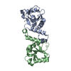
| ||||||||||||
|---|---|---|---|---|---|---|---|---|---|---|---|---|---|
| 1 | 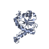
| ||||||||||||
| 2 | 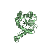
| ||||||||||||
| Unit cell |
|
-Components
| #1: Protein | Mass: 20025.602 Da / Num. of mol.: 2 Source method: isolated from a genetically manipulated source Source: (gene. exp.) Volvox reticuliferus (plant) / Gene: Vretifemale_1082 / Plasmid: pMAPLe4 / Production host:  #2: Water | ChemComp-HOH / |  Mass: 18.015 Da / Num. of mol.: 154 / Source method: isolated from a natural source / Formula: H2O Mass: 18.015 Da / Num. of mol.: 154 / Source method: isolated from a natural source / Formula: H2O |
|---|
-Experimental details
-Experiment
| Experiment | Method: X-RAY DIFFRACTION / Number of used crystals: 1 |
|---|
- Sample preparation
| Crystal | Density Matthews: 2.25 Å3/Da / Density % sol: 45.23 % |
|---|---|
| Crystal grow | Temperature: 293 K / Method: vapor diffusion, hanging drop Details: 0.2 M L-Proline, 0.1 M HEPES pH 7.5, 10% PEG3350. Cryoprotectant: 0.2 M L-Proline, 0.1 M HEPES pH 7.5, 23% PEG3350, 5% PEG400 |
-Data collection
| Diffraction | Mean temperature: 100 K / Serial crystal experiment: N |
|---|---|
| Diffraction source | Source: SYNCHROTRON / Site: APS / Beamline: 24-ID-C / Wavelength: 0.72932 Å |
| Detector | Type: DECTRIS EIGER2 X 16M / Detector: PIXEL / Date: Dec 14, 2022 |
| Radiation | Protocol: SINGLE WAVELENGTH / Monochromatic (M) / Laue (L): M / Scattering type: x-ray |
| Radiation wavelength | Wavelength: 0.72932 Å / Relative weight: 1 |
| Reflection | Resolution: 1.95→57.5 Å / Num. obs: 50761 / % possible obs: 99.88 % / Redundancy: 9.8 % / Biso Wilson estimate: 40.04 Å2 / CC1/2: 0.999 / CC star: 1 / Rmerge(I) obs: 0.08409 / Rpim(I) all: 0.02846 / Rrim(I) all: 0.08888 / Net I/σ(I): 14.03 |
| Reflection shell | Resolution: 1.95→2.02 Å / Redundancy: 10.1 % / Rmerge(I) obs: 0.8456 / Mean I/σ(I) obs: 1.77 / Num. unique obs: 2645 / CC1/2: 0.9 / CC star: 0.973 / Rpim(I) all: 0.2763 / Rrim(I) all: 0.8902 / % possible all: 99.77 |
- Processing
| Software |
| ||||||||||||||||||||||||||||||||||||||||||||||||||||||||||||||||||||||||||||||||||||||||||||||||||||||||||||||||||||||||||||||||||||||||||||
|---|---|---|---|---|---|---|---|---|---|---|---|---|---|---|---|---|---|---|---|---|---|---|---|---|---|---|---|---|---|---|---|---|---|---|---|---|---|---|---|---|---|---|---|---|---|---|---|---|---|---|---|---|---|---|---|---|---|---|---|---|---|---|---|---|---|---|---|---|---|---|---|---|---|---|---|---|---|---|---|---|---|---|---|---|---|---|---|---|---|---|---|---|---|---|---|---|---|---|---|---|---|---|---|---|---|---|---|---|---|---|---|---|---|---|---|---|---|---|---|---|---|---|---|---|---|---|---|---|---|---|---|---|---|---|---|---|---|---|---|---|---|
| Refinement | Method to determine structure: MOLECULAR REPLACEMENT / Resolution: 1.95→53.26 Å / SU ML: 0.2475 / Cross valid method: FREE R-VALUE / σ(F): 1.34 / Phase error: 25.3145 Stereochemistry target values: GeoStd + Monomer Library + CDL v1.2
| ||||||||||||||||||||||||||||||||||||||||||||||||||||||||||||||||||||||||||||||||||||||||||||||||||||||||||||||||||||||||||||||||||||||||||||
| Solvent computation | Shrinkage radii: 0.9 Å / VDW probe radii: 1.1 Å / Solvent model: FLAT BULK SOLVENT MODEL | ||||||||||||||||||||||||||||||||||||||||||||||||||||||||||||||||||||||||||||||||||||||||||||||||||||||||||||||||||||||||||||||||||||||||||||
| Displacement parameters | Biso mean: 51.15 Å2 | ||||||||||||||||||||||||||||||||||||||||||||||||||||||||||||||||||||||||||||||||||||||||||||||||||||||||||||||||||||||||||||||||||||||||||||
| Refinement step | Cycle: LAST / Resolution: 1.95→53.26 Å
| ||||||||||||||||||||||||||||||||||||||||||||||||||||||||||||||||||||||||||||||||||||||||||||||||||||||||||||||||||||||||||||||||||||||||||||
| Refine LS restraints |
| ||||||||||||||||||||||||||||||||||||||||||||||||||||||||||||||||||||||||||||||||||||||||||||||||||||||||||||||||||||||||||||||||||||||||||||
| LS refinement shell |
| ||||||||||||||||||||||||||||||||||||||||||||||||||||||||||||||||||||||||||||||||||||||||||||||||||||||||||||||||||||||||||||||||||||||||||||
| Refinement TLS params. | Method: refined / Origin x: -12.8609050956 Å / Origin y: -1.60037038005 Å / Origin z: 12.8250411556 Å
| ||||||||||||||||||||||||||||||||||||||||||||||||||||||||||||||||||||||||||||||||||||||||||||||||||||||||||||||||||||||||||||||||||||||||||||
| Refinement TLS group | Selection details: all |
