 Movie
Movie Controller

[English] 日本語
 Yorodumi
- PDB-8tf1: Crystal Structure of Pyridoxal Reductase (PDXI)in complex with NA... -
+ Open data

- Basic information
| Entry | Database: PDB / ID: 8tf1 | ||||||
|---|---|---|---|---|---|---|---|
| Title | Crystal Structure of Pyridoxal Reductase (PDXI)in complex with NADPH and Pyridoxal | ||||||
 Components Components | Pyridoxine 4-dehydrogenase | ||||||
 Keywords Keywords | OXIDOREDUCTASE / PDXI / Aldo-keto reductases superfamily / Vitamin B6 / NADPH / Pyridoxal | ||||||
| Function / homology |  Function and homology information Function and homology informationpyridoxine 4-dehydrogenase / pyridoxine 4-dehydrogenase (NADP+) activity / pyridoxal 5'-phosphate salvage / alcohol dehydrogenase (NADP+) activity / cytoplasm / cytosol Similarity search - Function | ||||||
| Biological species |  | ||||||
| Method |  X-RAY DIFFRACTION / MOLECULAR REPLACEMENT / Resolution: 2 Å X-RAY DIFFRACTION / MOLECULAR REPLACEMENT / Resolution: 2 Å | ||||||
 Authors Authors | Donkor, A.K. / Safo, M.K. / Musayev, F.N. | ||||||
| Funding support |  United States, 1items United States, 1items
| ||||||
 Citation Citation | Journal: Febs J. / Year: 2023 Title: Functional and structural properties of pyridoxal reductase (PdxI) from Escherichia coli: a pivotal enzyme in the vitamin B6 salvage pathway. Authors: Tramonti, A. / Donkor, A.K. / Parroni, A. / Musayev, F.N. / Barile, A. / Ghatge, M.S. / Graziani, C. / Alkhairi, M. / AlAwadh, M. / di Salvo, M.L. / Safo, M.K. / Contestabile, R. | ||||||
| History |
|
- Structure visualization
| Structure viewer | Molecule: MolmilJmol/JSmol |
|---|
- Downloads & links
-Download
| PDBx/mmCIF format | 8tf1.cif.gz | 80.3 KB | Display | PDBx/mmCIF format |
|---|---|---|---|---|
| PDB format | pdb8tf1.ent.gz | 56.8 KB | Display | PDB format |
| PDBx/mmJSON format | 8tf1.json.gz | Tree view | PDBx/mmJSON format | |
| Others |  Other downloads Other downloads |
-Validation report
| Arichive directory | https://data.pdbj.org/pub/pdb/validation_reports/tf/8tf1ftp://data.pdbj.org/pub/pdb/validation_reports/tf/8tf1 | HTTPS FTP |
|---|
-Related structure data


-Links
 PDBj
PDBj
- Assembly
| Deposited unit | 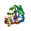
| ||||||||
|---|---|---|---|---|---|---|---|---|---|
| 1 |
| ||||||||
| Unit cell |
| ||||||||
| Components on special symmetry positions |
|
-Components
| #1: Protein | Mass: 32907.258 Da / Num. of mol.: 1 Source method: isolated from a genetically manipulated source Source: (gene. exp.) |
|---|---|
| #2: Chemical | ChemComp-NAP /   Mass: 743.405 Da / Num. of mol.: 1 / Source method: obtained synthetically / Formula: C21H28N7O17P3 Mass: 743.405 Da / Num. of mol.: 1 / Source method: obtained synthetically / Formula: C21H28N7O17P3 |
| #3: Chemical | ChemComp-UEG /   Mass: 169.178 Da / Num. of mol.: 1 / Source method: obtained synthetically / Formula: C8H11NO3 / Feature type: SUBJECT OF INVESTIGATION Mass: 169.178 Da / Num. of mol.: 1 / Source method: obtained synthetically / Formula: C8H11NO3 / Feature type: SUBJECT OF INVESTIGATION |
| #4: Chemical | ChemComp-MG /   Mass: 24.305 Da / Num. of mol.: 1 / Source method: obtained synthetically / Formula: Mg Mass: 24.305 Da / Num. of mol.: 1 / Source method: obtained synthetically / Formula: Mg |
| #5: Water | ChemComp-HOH /  Mass: 18.015 Da / Num. of mol.: 309 / Source method: isolated from a natural source / Formula: H2O Mass: 18.015 Da / Num. of mol.: 309 / Source method: isolated from a natural source / Formula: H2O |
| Has ligand of interest | Y |
| Has protein modification | N |
-Experimental details
-Experiment
| Experiment | Method: X-RAY DIFFRACTION / Number of used crystals: 1 |
|---|
- Sample preparation
| Crystal | Density Matthews: 2.28 Å3/Da / Density % sol: 46.12 % |
|---|---|
| Crystal grow | Temperature: 293 K / Method: vapor diffusion, sitting drop / pH: 6.5 / Details: 0.1M MES/NaOH, pH 6.5, 30% PEG400, 0.1M MgCl2 |
-Data collection
| Diffraction | Mean temperature: 100 K / Serial crystal experiment: N |
|---|---|
| Diffraction source | Source: ROTATING ANODE / Type: RIGAKU MICROMAX-007 HF / Wavelength: 1.5406 Å |
| Detector | Type: DECTRIS EIGER R 4M / Detector: PIXEL / Date: May 19, 2022 / Details: Rigaku VariMax-VHF Arc) Sec Confocal |
| Radiation | Protocol: SINGLE WAVELENGTH / Monochromatic (M) / Laue (L): M / Scattering type: x-ray |
| Radiation wavelength | Wavelength: 1.5406 Å / Relative weight: 1 |
| Reflection | Resolution: 2→27.18 Å / Num. obs: 21054 / % possible obs: 100 % / Redundancy: 7 % / CC1/2: 0.999 / Rmerge(I) obs: 0.068 / Rpim(I) all: 0.027 / Rrim(I) all: 0.073 / Net I/σ(I): 23.8 / Num. measured all: 147270 |
| Reflection shell | Resolution: 2→2.05 Å / % possible obs: 100 % / Redundancy: 6.9 % / Rmerge(I) obs: 0.443 / Num. measured all: 10752 / Num. unique obs: 1562 / CC1/2: 0.913 / Rpim(I) all: 0.18 / Rrim(I) all: 0.479 / Net I/σ(I) obs: 4.2 |
- Processing
| Software |
| ||||||||||||||||||||||||||||||||||||||||||||||||||||||||
|---|---|---|---|---|---|---|---|---|---|---|---|---|---|---|---|---|---|---|---|---|---|---|---|---|---|---|---|---|---|---|---|---|---|---|---|---|---|---|---|---|---|---|---|---|---|---|---|---|---|---|---|---|---|---|---|---|---|
| Refinement | Method to determine structure: MOLECULAR REPLACEMENT / Resolution: 2→27.177 Å / SU ML: 0.2 / Cross valid method: THROUGHOUT / σ(F): 1.35 / Phase error: 22.29 / Stereochemistry target values: ML
| ||||||||||||||||||||||||||||||||||||||||||||||||||||||||
| Solvent computation | Shrinkage radii: 0.9 Å / VDW probe radii: 1.11 Å / Solvent model: FLAT BULK SOLVENT MODEL | ||||||||||||||||||||||||||||||||||||||||||||||||||||||||
| Refinement step | Cycle: LAST / Resolution: 2→27.177 Å
| ||||||||||||||||||||||||||||||||||||||||||||||||||||||||
| Refine LS restraints |
| ||||||||||||||||||||||||||||||||||||||||||||||||||||||||
| LS refinement shell |
|
