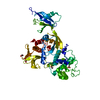
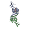
Entry Database : PDB / ID : 8tecTitle Crystal structure of Kindlin2 in complex with acylated beta1 integrin peptide Fermitin family homolog 2 Integrin beta-1 Keywords / / / Function / homology Function Domain/homology Component
/ / / / / / / / / / / / / / / / / / / / / / / / / / / / / / / / / / / / / / / / / / / / / / / / / / / / / / / / / / / / / / / / / / / / / / / / / / / / / / / / / / / / / / / / / / / / / / / / / / / / / / / / / / / / / / / / / / / / / / / / / / / / / / / / / / / / / / / / / / / / / / / / / / / / / / / / / / / / Biological species Mus musculus (house mouse)Method / / / Resolution : 2.04 Å Authors Zhang, P.F. / Wu, J.H. Funding support Organization Grant number Country National Institutes of Health/National Cancer Institute (NIH/NCI)
Journal : Iscience / Year : 2024Title : Acetyl-NPKY of integrin-beta 1 binds KINDLIN2 to control endothelial cell proliferation and junctional integrity.Authors : Sidibe, A. / Mykuliak, V.V. / Zhang, P. / Hytonen, V.P. / Wu, J. / Wehrle-Haller, B. History Deposition Jul 6, 2023 Deposition site / Processing site Revision 1.0 Jul 3, 2024 Provider / Type Revision 1.1 Oct 30, 2024 Group / Category / pdbx_modification_feature / Item
Show all Show less
 Movie
Movie Controller

 Yorodumi
 Open data
 Basic information
 Components
 Keywords
 Function and homology information
 X-RAY DIFFRACTION /
  Authors
 United States, 1items
  Citation
 Structure visualization
 Downloads & links
 Other downloads
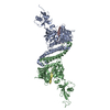
 PDBj
PDBj


 Assembly
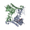

 Mass: 18.015 Da / Num. of mol.: 244 / Source method: isolated from a natural source / Formula: H2O
 Sample preparation
 Processing