 Movie
Movie Controller


+ Open data

- Basic information
| Entry | Database: PDB / ID: 8tcc | ||||||
|---|---|---|---|---|---|---|---|
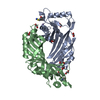
| Title | GTP Cyclohydrolase-IB with dehydrocostus lactone | ||||||
 Components Components | GTP cyclohydrolase FolE2 | ||||||
 Keywords Keywords | HYDROLASE/INHIBITOR / Cyclohydrolase / GTP / dehydrocostus lactone / fole2 / HYDROLASE-INHIBITOR complex | ||||||
| Function / homology |  Function and homology information Function and homology informationGTP cyclohydrolase I / GTP cyclohydrolase I activity / tetrahydrofolate biosynthetic process / metal ion binding Similarity search - Function | ||||||
| Biological species |  Burkholderia pseudomallei (bacteria) Burkholderia pseudomallei (bacteria) | ||||||
| Method |  X-RAY DIFFRACTION / SYNCHROTRON / MOLECULAR REPLACEMENT / Resolution: 3.1 Å X-RAY DIFFRACTION / SYNCHROTRON / MOLECULAR REPLACEMENT / Resolution: 3.1 Å | ||||||
 Authors Authors | McWhorter, K.L. / Amaya Lopez, C.Y. / Davis, K.M. | ||||||
| Funding support |  United States, 1items United States, 1items
| ||||||
 Citation Citation | Journal: Proc.Natl.Acad.Sci.USA / Year: 2024 Title: Combatting melioidosis with chemical synthetic lethality. Authors: Zhang, Y. / McWhorter, K.L. / Rosen, P.C. / Klaus, J.R. / Gallant, E. / Amaya Lopez, C.Y. / Jhunjhunwala, R. / Chandler, J.R. / Davis, K.M. / Seyedsayamdost, M.R. | ||||||
| History |
|
- Structure visualization
| Structure viewer | Molecule: MolmilJmol/JSmol |
|---|
- Downloads & links
-Download
| PDBx/mmCIF format | 8tcc.cif.gz | 207.3 KB | Display | PDBx/mmCIF format |
|---|---|---|---|---|
| PDB format | pdb8tcc.ent.gz | Display | PDB format | |
| PDBx/mmJSON format | 8tcc.json.gz | Tree view | PDBx/mmJSON format | |
| Others |  Other downloads Other downloads |
-Validation report
| Arichive directory | https://data.pdbj.org/pub/pdb/validation_reports/tc/8tccftp://data.pdbj.org/pub/pdb/validation_reports/tc/8tcc | HTTPS FTP |
|---|
-Related structure data
-Links
 PDBj
PDBj

- Assembly
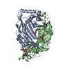
| Deposited unit | 
| ||||||||||||
|---|---|---|---|---|---|---|---|---|---|---|---|---|---|
| 1 |
| ||||||||||||
| Unit cell |
|
-Components
-Protein , 1 types, 4 molecules ABCD
| #1: Protein | Mass: 33811.340 Da / Num. of mol.: 4 Source method: isolated from a genetically manipulated source Source: (gene. exp.) Burkholderia pseudomallei (bacteria) / Gene: folE2 / Production host: |
|---|
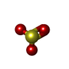
-Non-polymers , 5 types, 48 molecules 


| #2: Chemical | ChemComp-MN /  Mass: 54.938 Da / Num. of mol.: 1 / Source method: obtained synthetically / Formula: Mn Mass: 54.938 Da / Num. of mol.: 1 / Source method: obtained synthetically / Formula: Mn | ||||||
|---|---|---|---|---|---|---|---|
| #3: Chemical |  Mass: 62.068 Da / Num. of mol.: 2 / Source method: obtained synthetically / Formula: C2H6O2 Mass: 62.068 Da / Num. of mol.: 2 / Source method: obtained synthetically / Formula: C2H6O2#4: Chemical | ChemComp-SO3 / |  Mass: 80.063 Da / Num. of mol.: 1 / Source method: obtained synthetically / Formula: SO3 Mass: 80.063 Da / Num. of mol.: 1 / Source method: obtained synthetically / Formula: SO3#5: Chemical | Mass: 232.318 Da / Num. of mol.: 3 Source method: isolated from a genetically manipulated source Formula: C15H20O2 / Feature type: SUBJECT OF INVESTIGATION #6: Water | ChemComp-HOH / | Mass: 18.015 Da / Num. of mol.: 41 / Source method: isolated from a natural source / Formula: H2O |
-Details
| Has ligand of interest | Y |
|---|---|
| Has protein modification | Y |
-Experimental details
-Experiment
| Experiment | Method: X-RAY DIFFRACTION / Number of used crystals: 1 |
|---|
- Sample preparation
| Crystal | Density Matthews: 1.77 Å3/Da / Density % sol: 30.36 % |
|---|---|
| Crystal grow | Temperature: 293 K / Method: vapor diffusion, sitting drop Details: 0.1 M sodium chloride, 0.1 M Bis-Tris, 1.3-1.7 M ammonium sulfate, 0.01 M cadmium chloride PH range: 6.3-6.8 |
-Data collection
| Diffraction | Mean temperature: 100 K / Serial crystal experiment: N |
|---|---|
| Diffraction source | Source: SYNCHROTRON / Site: APS / Beamline: 21-ID-G / Wavelength: 0.97857 Å |
| Detector | Type: RAYONIX MX-300 / Detector: CCD / Date: Apr 13, 2022 |
| Radiation | Protocol: SINGLE WAVELENGTH / Monochromatic (M) / Laue (L): M / Scattering type: x-ray |
| Radiation wavelength | Wavelength: 0.97857 Å / Relative weight: 1 |
| Reflection | Resolution: 3.1→48.25 Å / Num. obs: 17590 / % possible obs: 96.85 % / Redundancy: 9.8 % / Biso Wilson estimate: 62.65 Å2 / CC1/2: 0.992 / CC star: 0.998 / Rmerge(I) obs: 0.2474 / Rpim(I) all: 0.07916 / Rrim(I) all: 0.2606 / Net I/σ(I): 10.13 |
| Reflection shell | Resolution: 3.1→3.211 Å / Redundancy: 10 % / Rmerge(I) obs: 1.149 / Mean I/σ(I) obs: 1.89 / Num. unique obs: 1716 / CC1/2: 0.834 / CC star: 0.954 / Rpim(I) all: 0.3566 / Rrim(I) all: 1.206 / % possible all: 97.55 |
- Processing
| Software |
| |||||||||||||||||||||||||||||||||||||||||||||||||
|---|---|---|---|---|---|---|---|---|---|---|---|---|---|---|---|---|---|---|---|---|---|---|---|---|---|---|---|---|---|---|---|---|---|---|---|---|---|---|---|---|---|---|---|---|---|---|---|---|---|---|
| Refinement | Method to determine structure: MOLECULAR REPLACEMENT / Resolution: 3.1→47.16 Å / SU ML: 0.5291 / Cross valid method: FREE R-VALUE / σ(F): 1.34 / Phase error: 32.569 Stereochemistry target values: GeoStd + Monomer Library + CDL v1.2
| |||||||||||||||||||||||||||||||||||||||||||||||||
| Solvent computation | Shrinkage radii: 0.9 Å / VDW probe radii: 1.1 Å / Solvent model: FLAT BULK SOLVENT MODEL | |||||||||||||||||||||||||||||||||||||||||||||||||
| Displacement parameters | Biso mean: 50.62 Å2 | |||||||||||||||||||||||||||||||||||||||||||||||||
| Refinement step | Cycle: LAST / Resolution: 3.1→47.16 Å
| |||||||||||||||||||||||||||||||||||||||||||||||||
| Refine LS restraints |
| |||||||||||||||||||||||||||||||||||||||||||||||||
| LS refinement shell |
|
