 Movie
Movie Controller

[English] 日本語
 Yorodumi
- PDB-8tb4: Sequence specific (AATT) orientation of DAPI molecules at a uniqu... -
+ Open data

- Basic information
| Entry | Database: PDB / ID: 8tb4 | ||||||||||||
|---|---|---|---|---|---|---|---|---|---|---|---|---|---|
| Title | Sequence specific (AATT) orientation of DAPI molecules at a unique minor groove binding site (position2) within a self-assembled 3D DNA lattice (4x5) | ||||||||||||
 Components Components |
| ||||||||||||
 Keywords Keywords | DNA / Self-Assembly / DNA Nanotechnology / DNA Scaffold / Crystal Lattice / Minor Groove Binders / Netropsin / DAPI / Hoechst / ImPyPy / polyamide / host-guest | ||||||||||||
| Function / homology | 6-AMIDINE-2-(4-AMIDINO-PHENYL)INDOLE / DNA / DNA (> 10) Function and homology information Function and homology information | ||||||||||||
| Biological species | synthetic construct (others) | ||||||||||||
| Method |  X-RAY DIFFRACTION / SYNCHROTRON / MOLECULAR REPLACEMENT / Resolution: 3.11 Å X-RAY DIFFRACTION / SYNCHROTRON / MOLECULAR REPLACEMENT / Resolution: 3.11 Å | ||||||||||||
 Authors Authors | Simmons, C.R. / MacCulloch, T. / Stephanopoulos, N. / Yan, H. | ||||||||||||
| Funding support |  United States, 3items United States, 3items
| ||||||||||||
 Citation Citation | Journal: J.Am.Chem.Soc. / Year: 2023 Title: Site-Specific Arrangement and Structure Determination of Minor Groove Binding Molecules in Self-Assembled Three-Dimensional DNA Crystals. Authors: Simmons, C.R. / Buchberger, A. / Henry, S.J.W. / Novacek, A. / Fahmi, N.E. / MacCulloch, T. / Stephanopoulos, N. / Yan, H. | ||||||||||||
| History |
|
- Structure visualization
| Structure viewer | Molecule: MolmilJmol/JSmol |
|---|
- Downloads & links
-Download
| PDBx/mmCIF format | 8tb4.cif.gz | 37.3 KB | Display | PDBx/mmCIF format |
|---|---|---|---|---|
| PDB format | pdb8tb4.ent.gz | 23.3 KB | Display | PDB format |
| PDBx/mmJSON format | 8tb4.json.gz | Tree view | PDBx/mmJSON format | |
| Others |  Other downloads Other downloads |
-Validation report
| Summary document | 8tb4_validation.pdf.gz | 596.3 KB | Display | wwPDB validaton report |
|---|---|---|---|---|
| Full document | 8tb4_full_validation.pdf.gz | 601.8 KB | Display | |
| Data in XML | 8tb4_validation.xml.gz | 4.2 KB | Display | |
| Data in CIF | 8tb4_validation.cif.gz | 5.2 KB | Display | |
| Arichive directory | https://data.pdbj.org/pub/pdb/validation_reports/tb/8tb4ftp://data.pdbj.org/pub/pdb/validation_reports/tb/8tb4 | HTTPS FTP |
-Related structure data
| Related structure data |  8t7bC 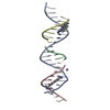 8t7xC  8t80C 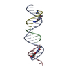 8ta8C  8ta9C  8tajC 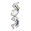 8tamC 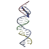 8tapC  8taqC  8tb3C  8tb8C  8tbdC  8tboC 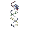 8tc2C  8tc4C  8tc6C  8tdtC C: citing same article ( |
|---|---|
| Similar structure data |
-Links
 PDBj
PDBj


- Assembly
| Deposited unit | 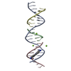
| ||||||||
|---|---|---|---|---|---|---|---|---|---|
| 1 |
| ||||||||
| Unit cell |
|
-Components
-DNA chain , 4 types, 4 molecules ABCD
| #1: DNA chain | Mass: 6465.213 Da / Num. of mol.: 1 / Source method: obtained synthetically / Source: (synth.) synthetic construct (others) |
|---|---|
| #2: DNA chain | Mass: 1480.012 Da / Num. of mol.: 1 / Source method: obtained synthetically / Source: (synth.) synthetic construct (others) |
| #3: DNA chain | Mass: 2720.808 Da / Num. of mol.: 1 / Source method: obtained synthetically / Source: (synth.) synthetic construct (others) |
| #4: DNA chain | Mass: 2129.409 Da / Num. of mol.: 1 / Source method: obtained synthetically / Source: (synth.) synthetic construct (others) |
-Non-polymers , 2 types, 4 molecules 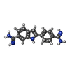

| #5: Chemical | ChemComp-DAP /  Mass: 277.324 Da / Num. of mol.: 1 / Source method: obtained synthetically / Formula: C16H15N5 Mass: 277.324 Da / Num. of mol.: 1 / Source method: obtained synthetically / Formula: C16H15N5 |
|---|---|
| #6: Chemical |  Mass: 24.305 Da / Num. of mol.: 3 / Source method: obtained synthetically / Formula: Mg Mass: 24.305 Da / Num. of mol.: 3 / Source method: obtained synthetically / Formula: Mg |
-Details
| Has ligand of interest | N |
|---|
-Experimental details
-Experiment
| Experiment | Method: X-RAY DIFFRACTION / Number of used crystals: 1 |
|---|
- Sample preparation
| Crystal | Density Matthews: 6.49 Å3/Da / Density % sol: 81.05 % |
|---|---|
| Crystal grow | Temperature: 298 K / Method: vapor diffusion, sitting drop Details: 0.5 mL of 0.05 M HEPES pH 7.5 with 15 mM MgCl2, 1.0 mM spermidine, and 10% dioxan was added to the reservoir with 2 uL added to the drop containing 4 uL of DNA stock. Temp details: temperature gradient generated from 60 to 25 C at 0.3 degrees per hour |
-Data collection
| Diffraction | Mean temperature: 100 K / Serial crystal experiment: N | ||||||||||||||||||||||||||||||||||||||||||||||||||||||||||||||||||||||||||||||||||||||||||||||||||||||||||||||||||||||||||||||||||||||||||||||||||||||||||||||||||||||||||||||||||||||||||||||||||||||||||||||||||
|---|---|---|---|---|---|---|---|---|---|---|---|---|---|---|---|---|---|---|---|---|---|---|---|---|---|---|---|---|---|---|---|---|---|---|---|---|---|---|---|---|---|---|---|---|---|---|---|---|---|---|---|---|---|---|---|---|---|---|---|---|---|---|---|---|---|---|---|---|---|---|---|---|---|---|---|---|---|---|---|---|---|---|---|---|---|---|---|---|---|---|---|---|---|---|---|---|---|---|---|---|---|---|---|---|---|---|---|---|---|---|---|---|---|---|---|---|---|---|---|---|---|---|---|---|---|---|---|---|---|---|---|---|---|---|---|---|---|---|---|---|---|---|---|---|---|---|---|---|---|---|---|---|---|---|---|---|---|---|---|---|---|---|---|---|---|---|---|---|---|---|---|---|---|---|---|---|---|---|---|---|---|---|---|---|---|---|---|---|---|---|---|---|---|---|---|---|---|---|---|---|---|---|---|---|---|---|---|---|---|---|---|
| Diffraction source | Source: SYNCHROTRON / Site: APS / Beamline: 19-ID / Wavelength: 0.92 Å | ||||||||||||||||||||||||||||||||||||||||||||||||||||||||||||||||||||||||||||||||||||||||||||||||||||||||||||||||||||||||||||||||||||||||||||||||||||||||||||||||||||||||||||||||||||||||||||||||||||||||||||||||||
| Detector | Type: DECTRIS PILATUS3 6M / Detector: PIXEL / Date: Sep 17, 2022 | ||||||||||||||||||||||||||||||||||||||||||||||||||||||||||||||||||||||||||||||||||||||||||||||||||||||||||||||||||||||||||||||||||||||||||||||||||||||||||||||||||||||||||||||||||||||||||||||||||||||||||||||||||
| Radiation | Protocol: SINGLE WAVELENGTH / Monochromatic (M) / Laue (L): M / Scattering type: x-ray | ||||||||||||||||||||||||||||||||||||||||||||||||||||||||||||||||||||||||||||||||||||||||||||||||||||||||||||||||||||||||||||||||||||||||||||||||||||||||||||||||||||||||||||||||||||||||||||||||||||||||||||||||||
| Radiation wavelength | Wavelength: 0.92 Å / Relative weight: 1 | ||||||||||||||||||||||||||||||||||||||||||||||||||||||||||||||||||||||||||||||||||||||||||||||||||||||||||||||||||||||||||||||||||||||||||||||||||||||||||||||||||||||||||||||||||||||||||||||||||||||||||||||||||
| Reflection | Resolution: 3.1→50 Å / Num. obs: 5774 / % possible obs: 99.7 % / Redundancy: 9.7 % / CC1/2: 1 / CC star: 1 / Rmerge(I) obs: 0.129 / Rpim(I) all: 0.042 / Rrim(I) all: 0.136 / Χ2: 4.546 / Net I/σ(I): 6.1 | ||||||||||||||||||||||||||||||||||||||||||||||||||||||||||||||||||||||||||||||||||||||||||||||||||||||||||||||||||||||||||||||||||||||||||||||||||||||||||||||||||||||||||||||||||||||||||||||||||||||||||||||||||
| Reflection shell | Diffraction-ID: 1
|
- Processing
| Software |
| ||||||||||||||||||||||||||||||||||||||||||||||||||||||||||||||||||||||||||||||||||||||||||||||||||||||||||||||||||||||||||||||||||||||||||||||||||||||||||||||||||||||||||||||||||||||
|---|---|---|---|---|---|---|---|---|---|---|---|---|---|---|---|---|---|---|---|---|---|---|---|---|---|---|---|---|---|---|---|---|---|---|---|---|---|---|---|---|---|---|---|---|---|---|---|---|---|---|---|---|---|---|---|---|---|---|---|---|---|---|---|---|---|---|---|---|---|---|---|---|---|---|---|---|---|---|---|---|---|---|---|---|---|---|---|---|---|---|---|---|---|---|---|---|---|---|---|---|---|---|---|---|---|---|---|---|---|---|---|---|---|---|---|---|---|---|---|---|---|---|---|---|---|---|---|---|---|---|---|---|---|---|---|---|---|---|---|---|---|---|---|---|---|---|---|---|---|---|---|---|---|---|---|---|---|---|---|---|---|---|---|---|---|---|---|---|---|---|---|---|---|---|---|---|---|---|---|---|---|---|---|
| Refinement | Method to determine structure: MOLECULAR REPLACEMENT / Resolution: 3.11→42.37 Å / Cor.coef. Fo:Fc: 0.981 / Cor.coef. Fo:Fc free: 0.962 / SU B: 20.035 / SU ML: 0.335 / Cross valid method: THROUGHOUT / ESU R: 0.433 / ESU R Free: 0.309 / Stereochemistry target values: MAXIMUM LIKELIHOOD / Details: HYDROGENS HAVE BEEN ADDED IN THE RIDING POSITIONS
| ||||||||||||||||||||||||||||||||||||||||||||||||||||||||||||||||||||||||||||||||||||||||||||||||||||||||||||||||||||||||||||||||||||||||||||||||||||||||||||||||||||||||||||||||||||||
| Solvent computation | Ion probe radii: 0.8 Å / Shrinkage radii: 0.8 Å / VDW probe radii: 1.2 Å / Solvent model: MASK | ||||||||||||||||||||||||||||||||||||||||||||||||||||||||||||||||||||||||||||||||||||||||||||||||||||||||||||||||||||||||||||||||||||||||||||||||||||||||||||||||||||||||||||||||||||||
| Displacement parameters | Biso mean: 138.397 Å2
| ||||||||||||||||||||||||||||||||||||||||||||||||||||||||||||||||||||||||||||||||||||||||||||||||||||||||||||||||||||||||||||||||||||||||||||||||||||||||||||||||||||||||||||||||||||||
| Refinement step | Cycle: 1 / Resolution: 3.11→42.37 Å
| ||||||||||||||||||||||||||||||||||||||||||||||||||||||||||||||||||||||||||||||||||||||||||||||||||||||||||||||||||||||||||||||||||||||||||||||||||||||||||||||||||||||||||||||||||||||
| Refine LS restraints |
|