 Movie
Movie Controller

[English] 日本語
 Yorodumi
- PDB-8tb2: Structure of SasG (type II) (residues 165-421) from Staphylococcu... -
+ Open data

- Basic information
| Entry | Database: PDB / ID: 8tb2 | |||||||||
|---|---|---|---|---|---|---|---|---|---|---|
| Title | Structure of SasG (type II) (residues 165-421) from Staphylococcus aureus MW2 | |||||||||
 Components Components | Putative surface protein MW2416 | |||||||||
 Keywords Keywords | CELL ADHESION / Lectin / SASG / adhesin | |||||||||
| Function / homology |  Function and homology information Function and homology information | |||||||||
| Biological species |  Staphylococcus aureus subsp. aureus MW2 (bacteria) Staphylococcus aureus subsp. aureus MW2 (bacteria) | |||||||||
| Method |  X-RAY DIFFRACTION / SYNCHROTRON / MOLECULAR REPLACEMENT / Resolution: 1.88 Å X-RAY DIFFRACTION / SYNCHROTRON / MOLECULAR REPLACEMENT / Resolution: 1.88 Å | |||||||||
 Authors Authors | Maciag, J.J. / Herr, A.B. | |||||||||
| Funding support |  United States, 2items United States, 2items
| |||||||||
 Citation Citation | Journal: Cell Rep / Year: 2024 Title: Staphylococcus aureus skin colonization is mediated by SasG lectin variation. Authors: Mills, K.B. / Maciag, J.J. / Wang, C. / Crawford, J.A. / Enroth, T.J. / Keim, K.C. / Dufrene, Y.F. / Robinson, D.A. / Fey, P.D. / Herr, A.B. / Horswill, A.R. | |||||||||
| History |
|
- Structure visualization
| Structure viewer | Molecule: MolmilJmol/JSmol |
|---|
- Downloads & links
-Download
| PDBx/mmCIF format | 8tb2.cif.gz | 68.7 KB | Display | PDBx/mmCIF format |
|---|---|---|---|---|
| PDB format | pdb8tb2.ent.gz | 48.4 KB | Display | PDB format |
| PDBx/mmJSON format | 8tb2.json.gz | Tree view | PDBx/mmJSON format | |
| Others |  Other downloads Other downloads |
-Validation report
| Summary document | 8tb2_validation.pdf.gz | 1.3 MB | Display | wwPDB validaton report |
|---|---|---|---|---|
| Full document | 8tb2_full_validation.pdf.gz | 1.3 MB | Display | |
| Data in XML | 8tb2_validation.xml.gz | 12.9 KB | Display | |
| Data in CIF | 8tb2_validation.cif.gz | 17.1 KB | Display | |
| Arichive directory | https://data.pdbj.org/pub/pdb/validation_reports/tb/8tb2ftp://data.pdbj.org/pub/pdb/validation_reports/tb/8tb2 | HTTPS FTP |
-Related structure data
| Similar structure data |
|---|
-Links
 PDBj
PDBj- Assembly
| Deposited unit | 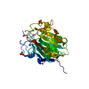
| ||||||||
|---|---|---|---|---|---|---|---|---|---|
| 1 |
| ||||||||
| Unit cell |
|
-Components
-Protein , 1 types, 1 molecules A
| #1: Protein | Mass: 28818.803 Da / Num. of mol.: 1 Source method: isolated from a genetically manipulated source Source: (gene. exp.) Staphylococcus aureus subsp. aureus MW2 (bacteria)Strain: MW2 / Gene: MW2416 / Production host: |
|---|
-Non-polymers , 5 types, 102 molecules 


| #2: Chemical | ChemComp-SO4 /  Mass: 96.063 Da / Num. of mol.: 4 / Source method: obtained synthetically / Formula: SO4 Mass: 96.063 Da / Num. of mol.: 4 / Source method: obtained synthetically / Formula: SO4#3: Chemical |  Mass: 40.078 Da / Num. of mol.: 2 / Source method: obtained synthetically / Formula: Ca / Feature type: SUBJECT OF INVESTIGATION Mass: 40.078 Da / Num. of mol.: 2 / Source method: obtained synthetically / Formula: Ca / Feature type: SUBJECT OF INVESTIGATION#4: Chemical | ChemComp-NA / |  Mass: 22.990 Da / Num. of mol.: 1 / Source method: obtained synthetically / Formula: Na Mass: 22.990 Da / Num. of mol.: 1 / Source method: obtained synthetically / Formula: Na#5: Chemical |  Mass: 65.409 Da / Num. of mol.: 3 / Source method: obtained synthetically / Formula: Zn Mass: 65.409 Da / Num. of mol.: 3 / Source method: obtained synthetically / Formula: Zn#6: Water | ChemComp-HOH / | Mass: 18.015 Da / Num. of mol.: 92 / Source method: isolated from a natural source / Formula: H2O |
|---|
-Details
| Has ligand of interest | Y |
|---|
-Experimental details
-Experiment
| Experiment | Method: X-RAY DIFFRACTION / Number of used crystals: 1 |
|---|
- Sample preparation
| Crystal | Density Matthews: 2.04 Å3/Da / Density % sol: 39.7 % |
|---|---|
| Crystal grow | Temperature: 298 K / Method: vapor diffusion, hanging drop / pH: 7.8 Details: 100 mM HEPES (pH 7.8), 200 mM Ammonium Sulfate, 25%-33% BCS PEG SMEAR Medium |
-Data collection
| Diffraction | Mean temperature: 100 K / Serial crystal experiment: N |
|---|---|
| Diffraction source | Source: SYNCHROTRON / Site: APS / Beamline: 24-ID-E / Wavelength: 0.9793384 Å |
| Detector | Type: DECTRIS EIGER X 16M / Detector: PIXEL / Date: Jun 16, 2021 |
| Radiation | Protocol: SINGLE WAVELENGTH / Monochromatic (M) / Laue (L): M / Scattering type: x-ray |
| Radiation wavelength | Wavelength: 0.9793384 Å / Relative weight: 1 |
| Reflection | Resolution: 1.88→52.36 Å / Num. obs: 20226 / % possible obs: 99.79 % / Redundancy: 2 % / Biso Wilson estimate: 20.27 Å2 / CC1/2: 0.991 / CC star: 0.998 / Rmerge(I) obs: 0.06672 / Rrim(I) all: 0.09436 / Net I/σ(I): 5.81 |
| Reflection shell | Resolution: 1.88→1.947 Å / Rmerge(I) obs: 0.2974 / Mean I/σ(I) obs: 2.25 / Num. unique obs: 1980 / CC1/2: 0.78 |
- Processing
| Software |
| ||||||||||||||||||||
|---|---|---|---|---|---|---|---|---|---|---|---|---|---|---|---|---|---|---|---|---|---|
| Refinement | Method to determine structure: MOLECULAR REPLACEMENT / Resolution: 1.88→52.36 Å / Cross valid method: THROUGHOUT
| ||||||||||||||||||||
| Refinement step | Cycle: LAST / Resolution: 1.88→52.36 Å
|
