 Movie
Movie Controller

[English] 日本語
 Yorodumi
- PDB-8t5j: Crystal structure of outer membrane lipoprotein carrier protein (... -
+ Open data

- Basic information
| Entry | Database: PDB / ID: 8t5j | ||||||
|---|---|---|---|---|---|---|---|
| Title | Crystal structure of outer membrane lipoprotein carrier protein (LolA) from Francisella philomiragia | ||||||
 Components Components | Outer membrane lipocarrier LolA family protein | ||||||
 Keywords Keywords | LIPID TRANSPORT / SSGCID / STRUCTURAL GENOMICS / SEATTLE STRUCTURAL GENOMICS CENTER FOR INFECTIOUS DISEASE / lipoprotein carrier / LolA | ||||||
| Function / homology | Outer membrane lipoprotein carrier protein LolA-like / Outer membrane lipoprotein carrier protein LolA-like / Lipoprotein localisation LolA/LolB/LppX / protein transport / metal ion binding / HEXANE-1,6-DIOL / Outer membrane lipocarrier LolA family protein Function and homology information Function and homology information | ||||||
| Biological species |  Francisella philomiragia (bacteria) Francisella philomiragia (bacteria) | ||||||
| Method |  X-RAY DIFFRACTION / MOLECULAR REPLACEMENT / Resolution: 1.55 Å X-RAY DIFFRACTION / MOLECULAR REPLACEMENT / Resolution: 1.55 Å | ||||||
 Authors Authors | Seattle Structural Genomics Center for Infectious Disease (SSGCID) | ||||||
| Funding support |  United States, 1items United States, 1items
| ||||||
 Citation Citation | Journal: To be published Title: Crystal structure of outer membrane lipoprotein carrier protein (LolA) from Francisella philomiragia Authors: Woodward, E.L. / Lovell, S. / Cooper, A. / Buchko, G. | ||||||
| History |
|
- Structure visualization
| Structure viewer | Molecule: MolmilJmol/JSmol |
|---|
- Downloads & links
-Download
| PDBx/mmCIF format | 8t5j.cif.gz | 86.6 KB | Display | PDBx/mmCIF format |
|---|---|---|---|---|
| PDB format | pdb8t5j.ent.gz | 63.2 KB | Display | PDB format |
| PDBx/mmJSON format | 8t5j.json.gz | Tree view | PDBx/mmJSON format | |
| Others |  Other downloads Other downloads |
-Validation report
| Arichive directory | https://data.pdbj.org/pub/pdb/validation_reports/t5/8t5jftp://data.pdbj.org/pub/pdb/validation_reports/t5/8t5j | HTTPS FTP |
|---|
-Related structure data
| Similar structure data |
|---|
-Links
 PDBj
PDBj- Assembly
| Deposited unit | 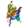
| |||||||||
|---|---|---|---|---|---|---|---|---|---|---|
| 1 |
| |||||||||
| Unit cell |
| |||||||||
| Components on special symmetry positions |
|
-Components
-Protein , 1 types, 1 molecules A
| #1: Protein | Mass: 22194.951 Da / Num. of mol.: 1 / Fragment: L32-N205 Source method: isolated from a genetically manipulated source Source: (gene. exp.) Francisella philomiragia (bacteria) / Gene: Fphi_1079 / Plasmid: FrphA.20731.a.VL2 / Production host: |
|---|
-Non-polymers , 5 types, 160 molecules 


| #2: Chemical |  Mass: 35.453 Da / Num. of mol.: 2 / Source method: obtained synthetically / Formula: Cl Mass: 35.453 Da / Num. of mol.: 2 / Source method: obtained synthetically / Formula: Cl#3: Chemical |  Mass: 24.305 Da / Num. of mol.: 2 / Source method: obtained synthetically / Formula: Mg Mass: 24.305 Da / Num. of mol.: 2 / Source method: obtained synthetically / Formula: Mg#4: Chemical |  Mass: 118.174 Da / Num. of mol.: 3 / Source method: obtained synthetically / Formula: C6H14O2 Mass: 118.174 Da / Num. of mol.: 3 / Source method: obtained synthetically / Formula: C6H14O2#5: Chemical | ChemComp-1PE / |  Mass: 238.278 Da / Num. of mol.: 1 / Source method: obtained synthetically / Formula: C10H22O6 / Comment: precipitant*YM Mass: 238.278 Da / Num. of mol.: 1 / Source method: obtained synthetically / Formula: C10H22O6 / Comment: precipitant*YM#6: Water | ChemComp-HOH / | Mass: 18.015 Da / Num. of mol.: 152 / Source method: isolated from a natural source / Formula: H2O |
|---|
-Details
| Has ligand of interest | N |
|---|
-Experimental details
-Experiment
| Experiment | Method: X-RAY DIFFRACTION / Number of used crystals: 1 |
|---|
- Sample preparation
| Crystal | Density Matthews: 2.15 Å3/Da / Density % sol: 42.91 % / Description: hexagonal prisms |
|---|---|
| Crystal grow | Temperature: 291 K / Method: vapor diffusion, sitting drop / pH: 7.5 Details: Berkeley D10: 20% (w/v) PEG 3350, 100 mM Hepes pH 7.5, 100 mM Lithium sulfate, 100 mM Magnesium chloride, 10% (w/v) 1,6-Hexanediol. FrphA.20731.a.VL2.PB00125 at 14.5 mg/mL. plate 13468 well ...Details: Berkeley D10: 20% (w/v) PEG 3350, 100 mM Hepes pH 7.5, 100 mM Lithium sulfate, 100 mM Magnesium chloride, 10% (w/v) 1,6-Hexanediol. FrphA.20731.a.VL2.PB00125 at 14.5 mg/mL. plate 13468 well D10 drop1, Cryo: 80% crystallant+20% PEG200 |
-Data collection
| Diffraction | Mean temperature: 100 K / Serial crystal experiment: N |
|---|---|
| Diffraction source | Source: SEALED TUBE / Type: BRUKER D8 QUEST / Wavelength: 1.5418 Å |
| Detector | Type: Bruker PHOTON III / Detector: PIXEL / Date: Jun 8, 2023 |
| Radiation | Monochromator: HELIOS MULTILAYER / Protocol: SINGLE WAVELENGTH / Monochromatic (M) / Laue (L): M / Scattering type: x-ray |
| Radiation wavelength | Wavelength: 1.5418 Å / Relative weight: 1 |
| Reflection | Resolution: 1.55→25.17 Å / Num. obs: 28420 / % possible obs: 100 % / Redundancy: 25.3 % / CC1/2: 1 / Rmerge(I) obs: 0.097 / Rpim(I) all: 0.02 / Rrim(I) all: 0.099 / Χ2: 0.94 / Net I/σ(I): 24.7 / Num. measured all: 718556 |
| Reflection shell | Resolution: 1.55→1.58 Å / % possible obs: 100 % / Redundancy: 17 % / Rmerge(I) obs: 2.648 / Num. measured all: 23447 / Num. unique obs: 1382 / CC1/2: 0.516 / Rpim(I) all: 0.662 / Rrim(I) all: 2.73 / Χ2: 1.35 / Net I/σ(I) obs: 1.6 |
- Processing
| Software |
| ||||||||||||||||||||||||||||||||||||||||||||||||||||||||||||||||||||||||||||||||||||||||||||||||||||||||||||||||||||||||||||||||||||||||||||||||||||||||||||||||||||||||||||||||||||||||||||||||||||||||
|---|---|---|---|---|---|---|---|---|---|---|---|---|---|---|---|---|---|---|---|---|---|---|---|---|---|---|---|---|---|---|---|---|---|---|---|---|---|---|---|---|---|---|---|---|---|---|---|---|---|---|---|---|---|---|---|---|---|---|---|---|---|---|---|---|---|---|---|---|---|---|---|---|---|---|---|---|---|---|---|---|---|---|---|---|---|---|---|---|---|---|---|---|---|---|---|---|---|---|---|---|---|---|---|---|---|---|---|---|---|---|---|---|---|---|---|---|---|---|---|---|---|---|---|---|---|---|---|---|---|---|---|---|---|---|---|---|---|---|---|---|---|---|---|---|---|---|---|---|---|---|---|---|---|---|---|---|---|---|---|---|---|---|---|---|---|---|---|---|---|---|---|---|---|---|---|---|---|---|---|---|---|---|---|---|---|---|---|---|---|---|---|---|---|---|---|---|---|---|---|---|---|
| Refinement | Method to determine structure: MOLECULAR REPLACEMENT / Resolution: 1.55→21.1 Å / SU ML: 0.15 / Cross valid method: FREE R-VALUE / σ(F): 1.04 / Phase error: 21.84 / Stereochemistry target values: ML
| ||||||||||||||||||||||||||||||||||||||||||||||||||||||||||||||||||||||||||||||||||||||||||||||||||||||||||||||||||||||||||||||||||||||||||||||||||||||||||||||||||||||||||||||||||||||||||||||||||||||||
| Solvent computation | Shrinkage radii: 0.9 Å / VDW probe radii: 1.1 Å / Solvent model: FLAT BULK SOLVENT MODEL | ||||||||||||||||||||||||||||||||||||||||||||||||||||||||||||||||||||||||||||||||||||||||||||||||||||||||||||||||||||||||||||||||||||||||||||||||||||||||||||||||||||||||||||||||||||||||||||||||||||||||
| Refinement step | Cycle: LAST / Resolution: 1.55→21.1 Å
| ||||||||||||||||||||||||||||||||||||||||||||||||||||||||||||||||||||||||||||||||||||||||||||||||||||||||||||||||||||||||||||||||||||||||||||||||||||||||||||||||||||||||||||||||||||||||||||||||||||||||
| Refine LS restraints |
| ||||||||||||||||||||||||||||||||||||||||||||||||||||||||||||||||||||||||||||||||||||||||||||||||||||||||||||||||||||||||||||||||||||||||||||||||||||||||||||||||||||||||||||||||||||||||||||||||||||||||
| LS refinement shell |
| ||||||||||||||||||||||||||||||||||||||||||||||||||||||||||||||||||||||||||||||||||||||||||||||||||||||||||||||||||||||||||||||||||||||||||||||||||||||||||||||||||||||||||||||||||||||||||||||||||||||||
| Refinement TLS params. | Method: refined / Refine-ID: X-RAY DIFFRACTION
| ||||||||||||||||||||||||||||||||||||||||||||||||||||||||||||||||||||||||||||||||||||||||||||||||||||||||||||||||||||||||||||||||||||||||||||||||||||||||||||||||||||||||||||||||||||||||||||||||||||||||
| Refinement TLS group |
|
