 Movie
Movie Controller

[English] 日本語
 Yorodumi
- PDB-8t5a: HIV-1 Integrase Catalytic Core Domain (CCD) F185H/Y99H/A128T Muta... -
+ Open data

- Basic information
| Entry | Database: PDB / ID: 8t5a | ||||||
|---|---|---|---|---|---|---|---|
| Title | HIV-1 Integrase Catalytic Core Domain (CCD) F185H/Y99H/A128T Mutant Complexed with STP03-0404 | ||||||
 Components Components | Integrase | ||||||
 Keywords Keywords | VIRAL PROTEIN / Integrase | ||||||
| Function / homology |  Function and homology information Function and homology informationexoribonuclease H activity / DNA integration / viral genome integration into host DNA / establishment of integrated proviral latency / RNA stem-loop binding / viral penetration into host nucleus / RNA-directed DNA polymerase activity / host cell / viral nucleocapsid / endonuclease activity ...exoribonuclease H activity / DNA integration / viral genome integration into host DNA / establishment of integrated proviral latency / RNA stem-loop binding / viral penetration into host nucleus / RNA-directed DNA polymerase activity / host cell / viral nucleocapsid / endonuclease activity / DNA recombination / aspartic-type endopeptidase activity / host cell cytoplasm / DNA-directed DNA polymerase activity / symbiont-mediated suppression of host gene expression / viral translational frameshifting / symbiont entry into host cell / lipid binding / host cell nucleus / host cell plasma membrane / proteolysis / DNA binding / zinc ion binding Similarity search - Function | ||||||
| Biological species |   Human immunodeficiency virus 1 Human immunodeficiency virus 1 | ||||||
| Method |  X-RAY DIFFRACTION / MOLECULAR REPLACEMENT / Resolution: 1.93 Å X-RAY DIFFRACTION / MOLECULAR REPLACEMENT / Resolution: 1.93 Å | ||||||
 Authors Authors | Dinh, T. / Kvaratskhelia, M. | ||||||
| Funding support |  United States, 1items United States, 1items
| ||||||
 Citation Citation | Journal: Biorxiv / Year: 2024 Title: The structural and mechanistic bases for the viral resistance to allosteric HIV-1 integrase inhibitor pirmitegravir. Authors: Dinh, T. / Tber, Z. / Rey, J.S. / Mengshetti, S. / Annamalai, A.S. / Haney, R. / Briganti, L. / Amblard, F. / Fuchs, J.R. / Cherepanov, P. / Kim, K. / Schinazi, R.F. / Perilla, J.R. / Kim, B. / Kvaratskhelia, M. | ||||||
| History |
|
- Structure visualization
| Structure viewer | Molecule: MolmilJmol/JSmol |
|---|
- Downloads & links
-Download
| PDBx/mmCIF format | 8t5a.cif.gz | 78.1 KB | Display | PDBx/mmCIF format |
|---|---|---|---|---|
| PDB format | pdb8t5a.ent.gz | 47.2 KB | Display | PDB format |
| PDBx/mmJSON format | 8t5a.json.gz | Tree view | PDBx/mmJSON format | |
| Others |  Other downloads Other downloads |
-Validation report
| Arichive directory | https://data.pdbj.org/pub/pdb/validation_reports/t5/8t5aftp://data.pdbj.org/pub/pdb/validation_reports/t5/8t5a | HTTPS FTP |
|---|
-Related structure data
| Related structure data |  8d3sC  8s9qSC 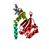 8t52C 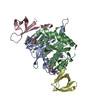 8t5bC S: Starting model for refinement C: citing same article ( |
|---|---|
| Similar structure data |
-Links
 PDBj
PDBj
- Assembly
| Deposited unit | 
| ||||||||||||
|---|---|---|---|---|---|---|---|---|---|---|---|---|---|
| 1 | 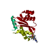
| ||||||||||||
| Unit cell |
|
-Components
| #1: Protein | Mass: 16770.176 Da / Num. of mol.: 1 / Mutation: F185H, Y99H, A128T Source method: isolated from a genetically manipulated source Source: (gene. exp.) Human immunodeficiency virus 1 / Gene: pol / Production host:  |
|---|---|
| #2: Chemical | ChemComp-WBV / (  Mass: 495.013 Da / Num. of mol.: 1 / Source method: obtained synthetically / Formula: C27H31ClN4O3 / Feature type: SUBJECT OF INVESTIGATION Mass: 495.013 Da / Num. of mol.: 1 / Source method: obtained synthetically / Formula: C27H31ClN4O3 / Feature type: SUBJECT OF INVESTIGATION |
| #3: Water | ChemComp-HOH /  Mass: 18.015 Da / Num. of mol.: 39 / Source method: isolated from a natural source / Formula: H2O Mass: 18.015 Da / Num. of mol.: 39 / Source method: isolated from a natural source / Formula: H2O |
| Has ligand of interest | Y |
-Experimental details
-Experiment
| Experiment | Method: X-RAY DIFFRACTION / Number of used crystals: 1 |
|---|
- Sample preparation
| Crystal | Density Matthews: 2.93 Å3/Da / Density % sol: 63.4 % |
|---|---|
| Crystal grow | Temperature: 277 K / Method: vapor diffusion, hanging drop / pH: 6.5 Details: 0.1 M (NH4)2SO4, 0.1 M sodium cacodylate (pH = 6.5), 10% PEG 8000, 5 mM DTT |
-Data collection
| Diffraction | Mean temperature: 100 K / Serial crystal experiment: N |
|---|---|
| Diffraction source | Source: ROTATING ANODE / Type: RIGAKU MICROMAX-007 HF / Wavelength: 1.5418 Å |
| Detector | Type: DECTRIS PILATUS 200K / Detector: PIXEL / Date: Dec 16, 2021 |
| Radiation | Protocol: SINGLE WAVELENGTH / Monochromatic (M) / Laue (L): M / Scattering type: x-ray |
| Radiation wavelength | Wavelength: 1.5418 Å / Relative weight: 1 |
| Reflection | Resolution: 1.93→45.28 Å / Num. obs: 10546 / % possible obs: 69.77 % / Redundancy: 2 % / Biso Wilson estimate: 25.2 Å2 / CC1/2: 0.993 / CC star: 0.998 / Rmerge(I) obs: 0.08091 / Rpim(I) all: 0.08091 / Rrim(I) all: 0.1144 / Net I/σ(I): 7.97 |
| Reflection shell | Resolution: 1.93→2.003 Å / Rmerge(I) obs: 0.3051 / Mean I/σ(I) obs: 1.64 / Num. unique obs: 39 / CC1/2: 0.788 / CC star: 0.939 / Rpim(I) all: 0.3051 / Rrim(I) all: 0.3051 / % possible all: 2.63 |
- Processing
| Software |
| |||||||||||||||||||||||||||||||||||
|---|---|---|---|---|---|---|---|---|---|---|---|---|---|---|---|---|---|---|---|---|---|---|---|---|---|---|---|---|---|---|---|---|---|---|---|---|
| Refinement | Method to determine structure: MOLECULAR REPLACEMENT Starting model: 8S9Q Resolution: 1.93→45.28 Å / SU ML: 0.2922 / Cross valid method: FREE R-VALUE / σ(F): 1.34 / Phase error: 34.7414 Stereochemistry target values: GeoStd + Monomer Library + CDL v1.2
| |||||||||||||||||||||||||||||||||||
| Solvent computation | Shrinkage radii: 0.9 Å / VDW probe radii: 1.11 Å / Solvent model: FLAT BULK SOLVENT MODEL | |||||||||||||||||||||||||||||||||||
| Displacement parameters | Biso mean: 31.44 Å2 | |||||||||||||||||||||||||||||||||||
| Refinement step | Cycle: LAST / Resolution: 1.93→45.28 Å
| |||||||||||||||||||||||||||||||||||
| Refine LS restraints |
| |||||||||||||||||||||||||||||||||||
| LS refinement shell |
|
