 Movie
Movie Controller

[English] 日本語
 Yorodumi
- PDB-8t2s: Structure of a group II intron ribonucleoprotein in the pre-branc... -
+ Open data

- Basic information
| Entry | Database: PDB / ID: 8t2s | ||||||
|---|---|---|---|---|---|---|---|
| Title | Structure of a group II intron ribonucleoprotein in the pre-branching (pre-1F) state | ||||||
 Components Components |
| ||||||
 Keywords Keywords | Transferase/RNA / RNP / RNA / Transferase-RNA complex | ||||||
| Function / homology |  Function and homology information Function and homology information | ||||||
| Biological species |  [Eubacterium] rectale (bacteria) [Eubacterium] rectale (bacteria) | ||||||
| Method | ELECTRON MICROSCOPY / single particle reconstruction / cryo EM / Resolution: 3 Å | ||||||
 Authors Authors | Xu, L. / Liu, T. / Chung, K. / Pyle, A.M. | ||||||
| Funding support |  United States, 1items United States, 1items
| ||||||
 Citation Citation | Journal: Nature / Year: 2023 Title: Structural insights into intron catalysis and dynamics during splicing. Authors: Ling Xu / Tianshuo Liu / Kevin Chung / Anna Marie Pyle / Abstract: The group II intron ribonucleoprotein is an archetypal splicing system with numerous mechanistic parallels to the spliceosome, including excision of lariat introns. Despite the importance of ...The group II intron ribonucleoprotein is an archetypal splicing system with numerous mechanistic parallels to the spliceosome, including excision of lariat introns. Despite the importance of branching in RNA metabolism, structural understanding of this process has remained elusive. Here we present a comprehensive analysis of three single-particle cryogenic electron microscopy structures captured along the splicing pathway. They reveal the network of molecular interactions that specifies the branchpoint adenosine and positions key functional groups to catalyse lariat formation and coordinate exon ligation. The structures also reveal conformational rearrangements of the branch helix and the mechanism of splice site exchange that facilitate the transition from branching to ligation. These findings shed light on the evolution of splicing and highlight the conservation of structural components, catalytic mechanism and dynamical strategies retained through time in premessenger RNA splicing machines. | ||||||
| History |
|
- Structure visualization
| Structure viewer | Molecule: MolmilJmol/JSmol |
|---|
- Downloads & links
-Download
| PDBx/mmCIF format | 8t2s.cif.gz | 394 KB | Display | PDBx/mmCIF format |
|---|---|---|---|---|
| PDB format | pdb8t2s.ent.gz | 297.3 KB | Display | PDB format |
| PDBx/mmJSON format | 8t2s.json.gz | Tree view | PDBx/mmJSON format | |
| Others |  Other downloads Other downloads |
-Validation report
| Arichive directory | https://data.pdbj.org/pub/pdb/validation_reports/t2/8t2sftp://data.pdbj.org/pub/pdb/validation_reports/t2/8t2s | HTTPS FTP |
|---|
-Related structure data
| Related structure data |  40986MC 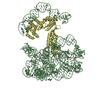 8t2rC 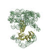 8t2tC M: map data used to model this data C: citing same article ( |
|---|---|
| Similar structure data |


-Links
 PDBj
PDBj


- Assembly
| Deposited unit | 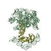
|
|---|---|
| 1 |
|
-Components
| #1: Protein | Mass: 49083.914 Da / Num. of mol.: 1 Source method: isolated from a genetically manipulated source Source: (gene. exp.) [Eubacterium] rectale (bacteria) / Gene: ltrA_2, ltrA, ERS852417_00966, FYL37_05080 / Production host: References: UniProt: A0A173ZME3, RNA-directed DNA polymerase | ||||
|---|---|---|---|---|---|
| #2: RNA chain | Mass: 211194.406 Da / Num. of mol.: 1 Source method: isolated from a genetically manipulated source Source: (gene. exp.) [Eubacterium] rectale (bacteria) / Production host: | ||||
| #3: Chemical | ChemComp-CA /   Mass: 40.078 Da / Num. of mol.: 11 / Source method: obtained synthetically / Formula: Ca / Feature type: SUBJECT OF INVESTIGATION Mass: 40.078 Da / Num. of mol.: 11 / Source method: obtained synthetically / Formula: Ca / Feature type: SUBJECT OF INVESTIGATION#4: Chemical |   Mass: 18.038 Da / Num. of mol.: 2 / Source method: obtained synthetically / Formula: H4N / Feature type: SUBJECT OF INVESTIGATION Mass: 18.038 Da / Num. of mol.: 2 / Source method: obtained synthetically / Formula: H4N / Feature type: SUBJECT OF INVESTIGATIONHas ligand of interest | Y | |
-Experimental details
-Experiment
| Experiment | Method: ELECTRON MICROSCOPY |
|---|---|
| EM experiment | Aggregation state: PARTICLE / 3D reconstruction method: single particle reconstruction |
- Sample preparation
| Component | Name: Intron RNP complex in pre-1f / Type: COMPLEX / Entity ID: #1-#2 / Source: RECOMBINANT |
|---|---|
| Molecular weight | Value: 0.3 MDa / Experimental value: NO |
| Source (natural) | Organism: [Eubacterium] rectale (bacteria) |
| Source (recombinant) | Organism: |
| Buffer solution | pH: 7.5 |
| Specimen | Embedding applied: NO / Shadowing applied: NO / Staining applied: NO / Vitrification applied: YES |
| Vitrification | Cryogen name: ETHANE |
- Electron microscopy imaging
| Experimental equipment |  Model: Titan Krios / Image courtesy: FEI Company |
|---|---|
| Microscopy | Model: FEI TITAN KRIOS |
| Electron gun | Electron source:  FIELD EMISSION GUN / Accelerating voltage: 300 kV / Illumination mode: FLOOD BEAM FIELD EMISSION GUN / Accelerating voltage: 300 kV / Illumination mode: FLOOD BEAM |
| Electron lens | Mode: DARK FIELD / Nominal defocus max: 2500 nm / Nominal defocus min: 1000 nm |
| Image recording | Electron dose: 1.5 e/Å2 / Film or detector model: GATAN K3 (6k x 4k) |
- Processing
| EM software | Name: PHENIX / Version: 1.20.1_4487: / Category: model refinement | ||||||||||||||||||||||||
|---|---|---|---|---|---|---|---|---|---|---|---|---|---|---|---|---|---|---|---|---|---|---|---|---|---|
| CTF correction | Type: NONE | ||||||||||||||||||||||||
| 3D reconstruction | Resolution: 3 Å / Resolution method: FSC 0.143 CUT-OFF / Num. of particles: 261619 / Symmetry type: POINT | ||||||||||||||||||||||||
| Refine LS restraints |
|
