[English] 日本語
Yorodumi
- PDB-8t2j: Structure of the catalytic domain of PPM1D/Wip1 serine/threonine ... -

+
Open data


ID or keywords:

Loading...

-
Basic information

Entry
Database: PDB / ID: 8t2j
TitleStructure of the catalytic domain of PPM1D/Wip1 serine/threonine phosphatase
ComponentsProtein phosphatase 1D
KeywordsHYDROLASE / Protein phosphatase / Metal-binding protein / Cell cycle / Cancer
Function / homology
Function and homology information


regulation of transcription initiation by RNA polymerase II / peptidyl-threonine dephosphorylation / Transcriptional regulation by RUNX2 / DNA methylation-dependent heterochromatin formation / mitogen-activated protein kinase binding / myosin phosphatase activity / negative regulation of gene expression, epigenetic / protein serine/threonine phosphatase activity / protein-serine/threonine phosphatase / DNA damage response, signal transduction by p53 class mediator ...regulation of transcription initiation by RNA polymerase II / peptidyl-threonine dephosphorylation / Transcriptional regulation by RUNX2 / DNA methylation-dependent heterochromatin formation / mitogen-activated protein kinase binding / myosin phosphatase activity / negative regulation of gene expression, epigenetic / protein serine/threonine phosphatase activity / protein-serine/threonine phosphatase / DNA damage response, signal transduction by p53 class mediator / cellular response to starvation / protein dephosphorylation / response to bacterium / response to radiation / G2/M transition of mitotic cell cycle / negative regulation of cell population proliferation / protein serine/threonine kinase activity / nucleolus / nucleoplasm / nucleus / metal ion binding / cytosol
Similarity search - Function
PPM-type phosphatase, divalent cation binding / PPM-type phosphatase domain signature. / Protein phosphatase 2C / Protein phosphatase 2C family / Serine/threonine phosphatases, family 2C, catalytic domain / PPM-type phosphatase domain profile. / PPM-type phosphatase-like domain / PPM-type phosphatase-like domain superfamily
Similarity search - Domain/homology
DI(HYDROXYETHYL)ETHER / Protein phosphatase 1D
Similarity search - Component
Biological speciesHomo sapiens (human)
MethodX-RAY DIFFRACTION / SYNCHROTRON / MOLECULAR REPLACEMENT / Resolution: 1.8 Å
AuthorsKumar, J.P. / Kosek, D. / Dyda, F.
Funding support United States, 1items
OrganizationGrant numberCountry
National Institutes of Health/National Institute of Diabetes and Digestive and Kidney Disease (NIH/NIDDK)Research resulting in this structure was funded in part by the Intramural Program of the NIDDK United States
CitationJournal: To Be Published
Title: Structure of the catalytic domain of PPM1D/Wip1 serine/threonine phosphatase
Authors: Kumar, J.P. / Kosek, D. / Dyda, F.
History
DepositionJun 6, 2023Deposition site: RCSB / Processing site: RCSB
Revision 1.0Jun 12, 2024Provider: repository / Type: Initial release

-
Structure visualization

Structure viewerMolecule:
MolmilJmol/JSmol

Downloads & links

-
Assembly

Deposited unit
A: Protein phosphatase 1D
hetero molecules


Theoretical massNumber of molelcules
Total (without water)38,6006
Polymers38,2611
Non-polymers3395
Water2,252125
1


  • Idetical with deposited unit
  • defined by author
  • Evidence: gel filtration
TypeNameSymmetry operationNumber
identity operation1_555x,y,z1
Unit cell
Length a, b, c (Å)49.664, 59.930, 49.566
Angle α, β, γ (deg.)90.000, 92.130, 90.000
Int Tables number4
Space group name H-MP1211
Space group name HallP2yb

-
Components

#1: Protein Protein phosphatase 1D / Protein phosphatase 2C isoform delta / PP2C-delta / Protein phosphatase magnesium-dependent 1 delta ...Protein phosphatase 2C isoform delta / PP2C-delta / Protein phosphatase magnesium-dependent 1 delta / p53-induced protein phosphatase 1


Mass: 38261.461 Da / Num. of mol.: 1 / Mutation: G8R, P33E, R250A, R258A, R259A
Source method: isolated from a genetically manipulated source
Source: (gene. exp.) Homo sapiens (human) / Gene: PPM1D, WIP1 / Plasmid: pE-SUMOstar Amp / Production host: Escherichia coli BL21(DE3) (bacteria) / Strain (production host): BL21(DE3) / Variant (production host): Rosetta
References: UniProt: O15297, protein-serine/threonine phosphatase
#2: Chemical ChemComp-MG / MAGNESIUM ION


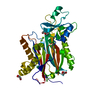
Mass: 24.305 Da / Num. of mol.: 2 / Source method: obtained synthetically / Formula: Mg
#3: Chemical ChemComp-PEG / DI(HYDROXYETHYL)ETHER


Mass: 106.120 Da / Num. of mol.: 1 / Source method: obtained synthetically / Formula: C4H10O3
#4: Chemical ChemComp-GOL / GLYCEROL / GLYCERIN / PROPANE-1,2,3-TRIOL


Mass: 92.094 Da / Num. of mol.: 2 / Source method: obtained synthetically / Formula: C3H8O3
#5: Water ChemComp-HOH / water


Mass: 18.015 Da / Num. of mol.: 125 / Source method: isolated from a natural source / Formula: H2O
Has ligand of interestY

-
Experimental details

-
Experiment

ExperimentMethod: X-RAY DIFFRACTION / Number of used crystals: 1

-
Sample preparation

CrystalDensity Matthews: 1.9 Å3/Da / Density % sol: 35.3 %
Crystal growTemperature: 298 K / Method: vapor diffusion, hanging drop / pH: 7.5
Details: 100 mM HEPES pH 7.5, 10% (w/v) PEG 3000, 20% PEG 400 (v/v), and 10% glycerol (v/v)

-
Data collection

DiffractionMean temperature: 100 K / Serial crystal experiment: N
Diffraction sourceSource: SYNCHROTRON / Site: APS / Beamline: 22-ID / Wavelength: 1 Å
DetectorType: DECTRIS EIGER X 16M / Detector: PIXEL / Date: Mar 20, 2020
RadiationProtocol: SINGLE WAVELENGTH / Monochromatic (M) / Laue (L): M / Scattering type: x-ray
Radiation wavelengthWavelength: 1 Å / Relative weight: 1
ReflectionResolution: 1.8→30 Å / Num. obs: 26792 / % possible obs: 98.9 % / Redundancy: 4.67 % / Biso Wilson estimate: 29.52 Å2 / CC1/2: 0.998 / Rmerge(I) obs: 0.066 / Rrim(I) all: 0.071 / Net I/σ(I): 15.75
Reflection shell
Resolution (Å)Rmerge(I) obsNum. unique obsCC1/2Rrim(I) allDiffraction-ID
1.8-1.850.49120100.9110.5321
1.85-1.90.37419110.9420.4041
1.9-1.950.31318550.9590.3371
1.95-2.010.23218170.9730.251
2.01-2.080.19117740.9790.2061
2.08-2.150.16317040.9810.1761
2.15-2.230.13116080.9860.1421
2.23-2.320.11215660.9930.121
2.32-2.430.09515390.9940.1021
2.43-2.550.08614730.9940.0931
2.55-2.680.07813970.9950.0841
2.68-2.850.07312980.9950.081
2.85-3.040.0712310.9950.0761
3.04-3.290.06511210.9950.0711
3.29-3.60.06210710.9950.0671
3.6-4.030.0579690.9960.0621
4.03-4.650.0558550.9970.061
4.65-5.690.0497200.9970.0541
5.69-8.050.0535570.9960.0581

-
Processing

Software
NameVersionClassification
PHENIX1.20.1_4487refinement
XSCALEdata scaling
XDSdata reduction
PHASERphasing
RefinementMethod to determine structure: MOLECULAR REPLACEMENT / Resolution: 1.8→25.65 Å / SU ML: 0.2131 / Cross valid method: FREE R-VALUE / σ(F): 2.02 / Phase error: 22.4976
Stereochemistry target values: GeoStd + Monomer Library + CDL v1.2
RfactorNum. reflection% reflection
Rfree0.2192 1072 4 %
Rwork0.1747 25718 -
obs0.1764 26790 98.9 %
Solvent computationShrinkage radii: 0.9 Å / VDW probe radii: 1.1 Å / Solvent model: FLAT BULK SOLVENT MODEL
Displacement parametersBiso mean: 37.69 Å2
Refinement stepCycle: LAST / Resolution: 1.8→25.65 Å
ProteinNucleic acidLigandSolventTotal
Num. atoms2635 0 21 125 2781
Refine LS restraints
Refine-IDTypeDev idealNumber
X-RAY DIFFRACTIONf_bond_d0.01462707
X-RAY DIFFRACTIONf_angle_d1.31593653
X-RAY DIFFRACTIONf_chiral_restr0.0891398
X-RAY DIFFRACTIONf_plane_restr0.0124471
X-RAY DIFFRACTIONf_dihedral_angle_d10.688372
LS refinement shell
Resolution (Å)Rfactor RfreeNum. reflection RfreeRfactor RworkNum. reflection RworkRefine-ID% reflection obs (%)
1.8-1.880.29641340.21853194X-RAY DIFFRACTION98.52
1.88-1.980.28361340.20083232X-RAY DIFFRACTION99.23
1.98-2.10.22861320.1833172X-RAY DIFFRACTION98.95
2.11-2.270.24351310.17583163X-RAY DIFFRACTION97.98
2.27-2.50.24451370.17723261X-RAY DIFFRACTION99.91
2.5-2.860.23251330.18653194X-RAY DIFFRACTION99.19
2.86-3.60.23761340.17963211X-RAY DIFFRACTION98.35
3.6-25.650.17971370.15993291X-RAY DIFFRACTION99.02

+
About Yorodumi

-
News

-
Feb 9, 2022. New format data for meta-information of EMDB entries

New format data for meta-information of EMDB entries

  • Version 3 of the EMDB header file is now the official format.
  • The previous official version 1.9 will be removed from the archive.

Related info.:EMDB header

External links:wwPDB to switch to version 3 of the EMDB data model

-
Aug 12, 2020. Covid-19 info

Covid-19 info

URL: https://pdbjlvh1.pdbj.org/emnavi/covid19.php

New page: Covid-19 featured information page in EM Navigator.

Related info.:Covid-19 info / Mar 5, 2020. Novel coronavirus structure data

+
Mar 5, 2020. Novel coronavirus structure data

Novel coronavirus structure data

Related info.:Yorodumi Speices / Aug 12, 2020. Covid-19 info

External links:COVID-19 featured content - PDBj / Molecule of the Month (242):Coronavirus Proteases

+
Jan 31, 2019. EMDB accession codes are about to change! (news from PDBe EMDB page)

EMDB accession codes are about to change! (news from PDBe EMDB page)

  • The allocation of 4 digits for EMDB accession codes will soon come to an end. Whilst these codes will remain in use, new EMDB accession codes will include an additional digit and will expand incrementally as the available range of codes is exhausted. The current 4-digit format prefixed with “EMD-” (i.e. EMD-XXXX) will advance to a 5-digit format (i.e. EMD-XXXXX), and so on. It is currently estimated that the 4-digit codes will be depleted around Spring 2019, at which point the 5-digit format will come into force.
  • The EM Navigator/Yorodumi systems omit the EMD- prefix.

Related info.:Q: What is EMD? / ID/Accession-code notation in Yorodumi/EM Navigator

External links:EMDB Accession Codes are Changing Soon! / Contact to PDBj

+
Jul 12, 2017. Major update of PDB

Major update of PDB

  • wwPDB released updated PDB data conforming to the new PDBx/mmCIF dictionary.
  • This is a major update changing the version number from 4 to 5, and with Remediation, in which all the entries are updated.
  • In this update, many items about electron microscopy experimental information are reorganized (e.g. em_software).
  • Now, EM Navigator and Yorodumi are based on the updated data.

External links:wwPDB Remediation / Enriched Model Files Conforming to OneDep Data Standards Now Available in the PDB FTP Archive

-
Yorodumi

Thousand views of thousand structures

  • Yorodumi is a browser for structure data from EMDB, PDB, SASBDB, etc.
  • This page is also the successor to EM Navigator detail page, and also detail information page/front-end page for Omokage search.
  • The word "yorodu" (or yorozu) is an old Japanese word meaning "ten thousand". "mi" (miru) is to see.

Related info.:EMDB / PDB / SASBDB / Comparison of 3 databanks / Yorodumi Search / Aug 31, 2016. New EM Navigator & Yorodumi / Yorodumi Papers / Jmol/JSmol / Function and homology information / Changes in new EM Navigator and Yorodumi

Read more