- PDB-8t0b: Novel Domain of Unknown Function Solved with AlphaFold -
+
データを開く
IDまたはキーワード:
読み込み中...
-
基本情報
登録情報
データベース: PDB / ID: 8t0b
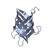
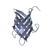
タイトル
Novel Domain of Unknown Function Solved with AlphaFold
要素
DUF1842 domain-containing protein
キーワード
UNKNOWN FUNCTION / AlphaFold / Bacterial Protein / Domain of Unknown Function / Novel Fold
機能・相同性
Domain of unknown function DUF1842 / Domain of unknown function DUF1843 / Domain of unknown function (DUF1842) / Domain of unknown function (DUF1843) / DUF1842 domain-containing protein
ジャーナル: Acta Crystallogr D Struct Biol / 年: 2024 タイトル: AlphaFold-assisted structure determination of a bacterial protein of unknown function using X-ray and electron crystallography. 著者: Justin E Miller / Matthew P Agdanowski / Joshua L Dolinsky / Michael R Sawaya / Duilio Cascio / Jose A Rodriguez / Todd O Yeates / 要旨: Macromolecular crystallography generally requires the recovery of missing phase information from diffraction data to reconstruct an electron-density map of the crystallized molecule. Most recent ...Macromolecular crystallography generally requires the recovery of missing phase information from diffraction data to reconstruct an electron-density map of the crystallized molecule. Most recent structures have been solved using molecular replacement as a phasing method, requiring an a priori structure that is closely related to the target protein to serve as a search model; when no such search model exists, molecular replacement is not possible. New advances in computational machine-learning methods, however, have resulted in major advances in protein structure predictions from sequence information. Methods that generate predicted structural models of sufficient accuracy provide a powerful approach to molecular replacement. Taking advantage of these advances, AlphaFold predictions were applied to enable structure determination of a bacterial protein of unknown function (UniProtKB Q63NT7, NCBI locus BPSS0212) based on diffraction data that had evaded phasing attempts using MIR and anomalous scattering methods. Using both X-ray and micro-electron (microED) diffraction data, it was possible to solve the structure of the main fragment of the protein using a predicted model of that domain as a starting point. The use of predicted structural models importantly expands the promise of electron diffraction, where structure determination relies critically on molecular replacement.
 ムービー
ムービー コントローラー


 データを開く
 基本情報
基本情報 要素
要素 キーワード
キーワード 機能・相同性情報
 Burkholderia pseudomallei (類鼻疽菌)
 X線回折 /
  データ登録者
 米国, 1件
  引用
引用 構造の表示
構造の表示 ダウンロードとリンク
 その他のダウンロード
 PDBj
PDBj 集合体
集合体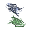

 分子量: 18.015 Da / 分子数: 11 / 由来タイプ: 天然 / 式: H2O
 試料調製
試料調製 解析
解析