- PDB-8t0b: Novel Domain of Unknown Function Solved with AlphaFold -
+
Open data
ID or keywords:
Loading...
-
Basic information
Entry
Database: PDB / ID: 8t0b
Title
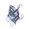
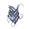
Novel Domain of Unknown Function Solved with AlphaFold
Components
DUF1842 domain-containing protein
Keywords
UNKNOWN FUNCTION / AlphaFold / Bacterial Protein / Domain of Unknown Function / Novel Fold
Function / homology
Domain of unknown function DUF1842 / Domain of unknown function DUF1843 / Domain of unknown function (DUF1842) / Domain of unknown function (DUF1843) / DUF1842 domain-containing protein
Journal: Acta Crystallogr D Struct Biol / Year: 2024 Title: AlphaFold-assisted structure determination of a bacterial protein of unknown function using X-ray and electron crystallography. Authors: Justin E Miller / Matthew P Agdanowski / Joshua L Dolinsky / Michael R Sawaya / Duilio Cascio / Jose A Rodriguez / Todd O Yeates / Abstract: Macromolecular crystallography generally requires the recovery of missing phase information from diffraction data to reconstruct an electron-density map of the crystallized molecule. Most recent ...Macromolecular crystallography generally requires the recovery of missing phase information from diffraction data to reconstruct an electron-density map of the crystallized molecule. Most recent structures have been solved using molecular replacement as a phasing method, requiring an a priori structure that is closely related to the target protein to serve as a search model; when no such search model exists, molecular replacement is not possible. New advances in computational machine-learning methods, however, have resulted in major advances in protein structure predictions from sequence information. Methods that generate predicted structural models of sufficient accuracy provide a powerful approach to molecular replacement. Taking advantage of these advances, AlphaFold predictions were applied to enable structure determination of a bacterial protein of unknown function (UniProtKB Q63NT7, NCBI locus BPSS0212) based on diffraction data that had evaded phasing attempts using MIR and anomalous scattering methods. Using both X-ray and micro-electron (microED) diffraction data, it was possible to solve the structure of the main fragment of the protein using a predicted model of that domain as a starting point. The use of predicted structural models importantly expands the promise of electron diffraction, where structure determination relies critically on molecular replacement.
In the structure databanks used in Yorodumi, some data are registered as the other names, "COVID-19 virus" and "2019-nCoV". Here are the details of the virus and the list of structure data.
Jan 31, 2019. EMDB accession codes are about to change! (news from PDBe EMDB page)
EMDB accession codes are about to change! (news from PDBe EMDB page)
The allocation of 4 digits for EMDB accession codes will soon come to an end. Whilst these codes will remain in use, new EMDB accession codes will include an additional digit and will expand incrementally as the available range of codes is exhausted. The current 4-digit format prefixed with “EMD-” (i.e. EMD-XXXX) will advance to a 5-digit format (i.e. EMD-XXXXX), and so on. It is currently estimated that the 4-digit codes will be depleted around Spring 2019, at which point the 5-digit format will come into force.
The EM Navigator/Yorodumi systems omit the EMD- prefix.
Related info.:Q: What is EMD? / ID/Accession-code notation in Yorodumi/EM Navigator
Yorodumi is a browser for structure data from EMDB, PDB, SASBDB, etc.
This page is also the successor to EM Navigator detail page, and also detail information page/front-end page for Omokage search.
The word "yorodu" (or yorozu) is an old Japanese word meaning "ten thousand". "mi" (miru) is to see.
Related info.:EMDB / PDB / SASBDB / Comparison of 3 databanks / Yorodumi Search / Aug 31, 2016. New EM Navigator & Yorodumi / Yorodumi Papers / Jmol/JSmol / Function and homology information / Changes in new EM Navigator and Yorodumi
 Movie
Movie Controller


 Open data
 Basic information
 Components
 Keywords
 Function and homology information
 Burkholderia pseudomallei (bacteria)
 X-RAY DIFFRACTION /
  Authors
 United States, 1items
  Citation
 Structure visualization
 Downloads & links
 Other downloads
 PDBj
PDBj Assembly

 Mass: 18.015 Da / Num. of mol.: 11 / Source method: isolated from a natural source / Formula: H2O
 Sample preparation
 Processing