 Movie
Movie Controller


+ Open data

- Basic information
| Entry | Database: PDB / ID: 8szo | |||||||||
|---|---|---|---|---|---|---|---|---|---|---|
| Title | Canavalia villosa lectin in complex with alpha-methyl-mannoside | |||||||||
 Components Components | Canavalia villosa lectin | |||||||||
 Keywords Keywords | SUGAR BINDING PROTEIN / Carbohydrate binding protein / Plant lectin / beta-sandwich / pro-inflammatory / PLANT PROTEIN | |||||||||
| Function / homology | methyl alpha-D-mannopyranoside / :  Function and homology information Function and homology information | |||||||||
| Biological species |  Canavalia villosa (plant) Canavalia villosa (plant) | |||||||||
| Method |  X-RAY DIFFRACTION / SYNCHROTRON / MOLECULAR REPLACEMENT / Resolution: 2.5 Å X-RAY DIFFRACTION / SYNCHROTRON / MOLECULAR REPLACEMENT / Resolution: 2.5 Å | |||||||||
 Authors Authors | Cavada, B.S. / Lossio, C.F. / Pinto-Junior, V.R. / Osterne, V.J.S. / Oliveira, M.V. / Neco, A.H.B. / Nascimento, K.S. | |||||||||
| Funding support |  Brazil, 2items Brazil, 2items
| |||||||||
 Citation Citation | Journal: Int J Biol Macromol / Year: 2017 Title: Lectin from Canavalia villosa seeds: A glucose/mannose-specific protein and a new tool for inflammation studies. Authors: Lossio, C.F. / Moreira, C.G. / Amorim, R.M.F. / Nobre, C.S. / Silva, M.T.L. / Neto, C.C. / Pinto-Junior, V.R. / Silva, I.B. / Campos, J. / Assreuy, A.M.S. / Cavada, B.S. / Nascimento, K.S. | |||||||||
| History |
|
- Structure visualization
| Structure viewer | Molecule: MolmilJmol/JSmol |
|---|
- Downloads & links
-Download
| PDBx/mmCIF format | 8szo.cif.gz | 110.4 KB | Display | PDBx/mmCIF format |
|---|---|---|---|---|
| PDB format | pdb8szo.ent.gz | 82.5 KB | Display | PDB format |
| PDBx/mmJSON format | 8szo.json.gz | Tree view | PDBx/mmJSON format | |
| Others |  Other downloads Other downloads |
-Validation report
| Arichive directory | https://data.pdbj.org/pub/pdb/validation_reports/sz/8szoftp://data.pdbj.org/pub/pdb/validation_reports/sz/8szo | HTTPS FTP |
|---|
-Related structure data
| Similar structure data |
|---|
-Links
 PDBj
PDBj- Assembly
| Deposited unit | 
| ||||||||
|---|---|---|---|---|---|---|---|---|---|
| 1 | 
| ||||||||
| Unit cell |
|
-Components
-Protein / Sugars , 2 types, 2 molecules A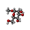
| #1: Protein | Mass: 25685.461 Da / Num. of mol.: 1 / Source method: isolated from a natural source Details: The residues S117, N118, S119 and T120 in the coordinate sequence are located in a loop region of the tertiary structure and were not detected due to lack of electron density on this ...Details: The residues S117, N118, S119 and T120 in the coordinate sequence are located in a loop region of the tertiary structure and were not detected due to lack of electron density on this specific region. This data deficiency is frequently observed in other ConA-like lectins structures solved by macromolecular crystallography. The sample sequence displays the full 237 amino acid of the protein. Source: (natural) Canavalia villosa (plant) |
|---|---|
| #4: Sugar | ChemComp-MMA /  Type: D-saccharide / Mass: 194.182 Da / Num. of mol.: 1 / Source method: obtained synthetically / Formula: C7H14O6 / Feature type: SUBJECT OF INVESTIGATION Type: D-saccharide / Mass: 194.182 Da / Num. of mol.: 1 / Source method: obtained synthetically / Formula: C7H14O6 / Feature type: SUBJECT OF INVESTIGATION |
-Non-polymers , 4 types, 91 molecules 


| #2: Chemical | ChemComp-MN /  Mass: 54.938 Da / Num. of mol.: 1 / Source method: obtained synthetically / Formula: Mn / Feature type: SUBJECT OF INVESTIGATION Mass: 54.938 Da / Num. of mol.: 1 / Source method: obtained synthetically / Formula: Mn / Feature type: SUBJECT OF INVESTIGATION |
|---|---|
| #3: Chemical | ChemComp-CA /  Mass: 40.078 Da / Num. of mol.: 1 / Source method: obtained synthetically / Formula: Ca / Feature type: SUBJECT OF INVESTIGATION Mass: 40.078 Da / Num. of mol.: 1 / Source method: obtained synthetically / Formula: Ca / Feature type: SUBJECT OF INVESTIGATION |
| #5: Chemical | ChemComp-GOL /  Mass: 92.094 Da / Num. of mol.: 1 / Source method: isolated from a natural source / Formula: C3H8O3 Mass: 92.094 Da / Num. of mol.: 1 / Source method: isolated from a natural source / Formula: C3H8O3 |
| #6: Water | ChemComp-HOH / Mass: 18.015 Da / Num. of mol.: 88 / Source method: isolated from a natural source / Formula: H2O |
-Details
| Has ligand of interest | Y |
|---|
-Experimental details
-Experiment
| Experiment | Method: X-RAY DIFFRACTION / Number of used crystals: 1 |
|---|
- Sample preparation
| Crystal | Density Matthews: 2.25 Å3/Da / Density % sol: 45.35 % |
|---|---|
| Crystal grow | Temperature: 298 K / Method: vapor diffusion / pH: 4.2 Details: PEG1000, potassium citrate phosphate, lithium sulfate |
-Data collection
| Diffraction | Mean temperature: 100 K / Serial crystal experiment: N |
|---|---|
| Diffraction source | Source: SYNCHROTRON / Site: LNLS / Beamline: W01B-MX2 / Wavelength: 1.45 Å |
| Detector | Type: DECTRIS PILATUS 2M / Detector: PIXEL / Date: Oct 9, 2017 |
| Radiation | Protocol: SINGLE WAVELENGTH / Monochromatic (M) / Laue (L): M / Scattering type: x-ray |
| Radiation wavelength | Wavelength: 1.45 Å / Relative weight: 1 |
| Reflection | Resolution: 2.5→27.48 Å / Num. obs: 8301 / % possible obs: 99.8 % / Redundancy: 4.9 % / CC1/2: 0.991 / Rmerge(I) obs: 0.112 / Rpim(I) all: 0.056 / Rrim(I) all: 0.126 / Net I/σ(I): 8.9 / Num. measured all: 40674 |
| Reflection shell | Resolution: 2.5→2.64 Å / % possible obs: 99.7 % / Redundancy: 4.5 % / Rmerge(I) obs: 0.433 / Num. measured all: 5399 / Num. unique obs: 1200 / CC1/2: 0.867 / Rpim(I) all: 0.224 / Rrim(I) all: 0.489 / Net I/σ(I) obs: 3 |
- Processing
| Software |
| ||||||||||||||||||||||||||||||||||||||||
|---|---|---|---|---|---|---|---|---|---|---|---|---|---|---|---|---|---|---|---|---|---|---|---|---|---|---|---|---|---|---|---|---|---|---|---|---|---|---|---|---|---|
| Refinement | Method to determine structure: MOLECULAR REPLACEMENT / Resolution: 2.5→27.48 Å / SU ML: 0.28 / Cross valid method: FREE R-VALUE / σ(F): 1.34 / Phase error: 28.37 / Stereochemistry target values: ML
| ||||||||||||||||||||||||||||||||||||||||
| Solvent computation | Shrinkage radii: 0.9 Å / VDW probe radii: 1.1 Å / Solvent model: FLAT BULK SOLVENT MODEL | ||||||||||||||||||||||||||||||||||||||||
| Refinement step | Cycle: LAST / Resolution: 2.5→27.48 Å
| ||||||||||||||||||||||||||||||||||||||||
| Refine LS restraints |
| ||||||||||||||||||||||||||||||||||||||||
| LS refinement shell |
| ||||||||||||||||||||||||||||||||||||||||
| Refinement TLS params. | Method: refined / Origin x: 14.6292 Å / Origin y: 14.4776 Å / Origin z: 11.3444 Å
| ||||||||||||||||||||||||||||||||||||||||
| Refinement TLS group | Selection details: all |
