 Movie
Movie Controller

[English] 日本語
 Yorodumi
- PDB-8sya: X-ray crystal structure of UDP-2,3-diacetamido-2,3-dideoxy-glucur... -
+ Open data

- Basic information
| Entry | Database: PDB / ID: 8sya | ||||||
|---|---|---|---|---|---|---|---|
| Title | X-ray crystal structure of UDP-2,3-diacetamido-2,3-dideoxy-glucuronic acid-2-epimerase from Thermus thermophilus strain HB27, D98N variant in the presence of UDP-2,3-diacetamido-2,3-dideoxy-glucuronic acid and UDP at pH 9 | ||||||
 Components Components | UDP-2,3-diacetamido-2,3-dideoxy-glucuronic acid-2-epimerase | ||||||
 Keywords Keywords | ISOMERASE / lipopolysaccharide / O-antigen / 2-epimerase | ||||||
| Function / homology |  Function and homology information Function and homology informationUDP-N-acetylglucosamine 2-epimerase (non-hydrolysing) / UDP-N-acetylglucosamine 2-epimerase activity / nucleotide binding Similarity search - Function | ||||||
| Biological species |   Thermus thermophilus HB27 (bacteria) Thermus thermophilus HB27 (bacteria) | ||||||
| Method |  X-RAY DIFFRACTION / FOURIER SYNTHESIS / Resolution: 2.3 Å X-RAY DIFFRACTION / FOURIER SYNTHESIS / Resolution: 2.3 Å | ||||||
 Authors Authors | Thoden, J.B. / Holden, H.M. | ||||||
| Funding support |  United States, 1items United States, 1items
| ||||||
 Citation Citation | Journal: J.Biol.Chem. / Year: 2023 Title: Structural analysis of a bacterial UDP-sugar 2-epimerase reveals the active site architecture before and after catalysis. Authors: Thoden, J.B. / McKnight, J.O. / Kroft, C.W. / Jast, J.D.T. / Holden, H.M. | ||||||
| History |
|
- Structure visualization
| Structure viewer | Molecule: MolmilJmol/JSmol |
|---|
- Downloads & links
-Download
| PDBx/mmCIF format | 8sya.cif.gz | 161 KB | Display | PDBx/mmCIF format |
|---|---|---|---|---|
| PDB format | pdb8sya.ent.gz | 124.9 KB | Display | PDB format |
| PDBx/mmJSON format | 8sya.json.gz | Tree view | PDBx/mmJSON format | |
| Others |  Other downloads Other downloads |
-Validation report
| Arichive directory | https://data.pdbj.org/pub/pdb/validation_reports/sy/8syaftp://data.pdbj.org/pub/pdb/validation_reports/sy/8sya | HTTPS FTP |
|---|
-Related structure data
| Related structure data |  8sxvC  8sxwC  8sxyC  8sy0C  8sy9C  8sybC  8sydC  8syeC 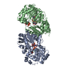 8syhC C: citing same article ( |
|---|---|
| Similar structure data |
-Links
 PDBj
PDBj


- Assembly
| Deposited unit | 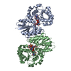
| ||||||||
|---|---|---|---|---|---|---|---|---|---|
| 1 |
| ||||||||
| Unit cell |
|
-Components
| #1: Protein | Mass: 41994.086 Da / Num. of mol.: 2 / Mutation: D98N Source method: isolated from a genetically manipulated source Source: (gene. exp.) Thermus thermophilus HB27 (bacteria) / Strain: ATCC BAA-163 / DSM 7039 / HB27 / Gene: TT_C0285 / Production host: #2: Chemical |   Mass: 662.389 Da / Num. of mol.: 2 / Source method: obtained synthetically / Formula: C19H28N4O18P2 / Feature type: SUBJECT OF INVESTIGATION Mass: 662.389 Da / Num. of mol.: 2 / Source method: obtained synthetically / Formula: C19H28N4O18P2 / Feature type: SUBJECT OF INVESTIGATION#3: Chemical | ChemComp-UDP / |   Type: RNA linking / Mass: 404.161 Da / Num. of mol.: 1 / Source method: obtained synthetically / Formula: C9H14N2O12P2 / Feature type: SUBJECT OF INVESTIGATION / Comment: UDP*YM Type: RNA linking / Mass: 404.161 Da / Num. of mol.: 1 / Source method: obtained synthetically / Formula: C9H14N2O12P2 / Feature type: SUBJECT OF INVESTIGATION / Comment: UDP*YM#4: Water | ChemComp-HOH / |  Mass: 18.015 Da / Num. of mol.: 151 / Source method: isolated from a natural source / Formula: H2O Mass: 18.015 Da / Num. of mol.: 151 / Source method: isolated from a natural source / Formula: H2OHas ligand of interest | Y | |
|---|
-Experimental details
-Experiment
| Experiment | Method: X-RAY DIFFRACTION / Number of used crystals: 1 |
|---|
- Sample preparation
| Crystal | Density Matthews: 2.35 Å3/Da / Density % sol: 47.63 % |
|---|---|
| Crystal grow | Temperature: 293 K / Method: vapor diffusion, hanging drop / pH: 9 Details: Protein incubated with 5 mM UDP-2,3-diacetamido-2,3-dideoxy-glucuronic acid. Precipitant used was 18-22% poly(ethylene glycol) 8000, 100 mM CHES (pH 9) |
-Data collection
| Diffraction | Mean temperature: 100 K / Serial crystal experiment: N |
|---|---|
| Diffraction source | Source: SEALED TUBE / Type: BRUKER D8 QUEST / Wavelength: 1.5418 Å |
| Detector | Type: Bruker PHOTON II / Detector: PIXEL / Date: Mar 20, 2023 |
| Radiation | Protocol: SINGLE WAVELENGTH / Monochromatic (M) / Laue (L): M / Scattering type: x-ray |
| Radiation wavelength | Wavelength: 1.5418 Å / Relative weight: 1 |
| Reflection | Resolution: 2.3→50 Å / Num. obs: 34302 / % possible obs: 99.6 % / Observed criterion σ(F): 0 / Observed criterion σ(I): 0 / Redundancy: 8.5 % / Rsym value: 0.096 / Net I/σ(I): 9.7 |
| Reflection shell | Resolution: 2.3→2.4 Å / Redundancy: 3.8 % / Num. unique obs: 4050 / R split: 2.2 / Rsym value: 0.45 / % possible all: 98.8 |
- Processing
| Software |
| ||||||||||||||||||||||||||||||||||||||||||||||||||||||||||||||||||||||||||||||||||||||||||||||||||||||||||||||||||||||||||||||||||||||||||||||||||||||||||||||||||||||||||||||||||||||
|---|---|---|---|---|---|---|---|---|---|---|---|---|---|---|---|---|---|---|---|---|---|---|---|---|---|---|---|---|---|---|---|---|---|---|---|---|---|---|---|---|---|---|---|---|---|---|---|---|---|---|---|---|---|---|---|---|---|---|---|---|---|---|---|---|---|---|---|---|---|---|---|---|---|---|---|---|---|---|---|---|---|---|---|---|---|---|---|---|---|---|---|---|---|---|---|---|---|---|---|---|---|---|---|---|---|---|---|---|---|---|---|---|---|---|---|---|---|---|---|---|---|---|---|---|---|---|---|---|---|---|---|---|---|---|---|---|---|---|---|---|---|---|---|---|---|---|---|---|---|---|---|---|---|---|---|---|---|---|---|---|---|---|---|---|---|---|---|---|---|---|---|---|---|---|---|---|---|---|---|---|---|---|---|
| Refinement | Method to determine structure: FOURIER SYNTHESIS / Resolution: 2.3→44.79 Å / Cor.coef. Fo:Fc: 0.931 / Cor.coef. Fo:Fc free: 0.902 / SU B: 10.903 / SU ML: 0.232 / Cross valid method: THROUGHOUT / ESU R: 0.387 / ESU R Free: 0.243 / Stereochemistry target values: MAXIMUM LIKELIHOOD / Details: HYDROGENS HAVE BEEN ADDED IN THE RIDING POSITIONS
| ||||||||||||||||||||||||||||||||||||||||||||||||||||||||||||||||||||||||||||||||||||||||||||||||||||||||||||||||||||||||||||||||||||||||||||||||||||||||||||||||||||||||||||||||||||||
| Solvent computation | Ion probe radii: 0.8 Å / Shrinkage radii: 0.8 Å / VDW probe radii: 1.2 Å / Solvent model: MASK | ||||||||||||||||||||||||||||||||||||||||||||||||||||||||||||||||||||||||||||||||||||||||||||||||||||||||||||||||||||||||||||||||||||||||||||||||||||||||||||||||||||||||||||||||||||||
| Displacement parameters | Biso mean: 29.641 Å2
| ||||||||||||||||||||||||||||||||||||||||||||||||||||||||||||||||||||||||||||||||||||||||||||||||||||||||||||||||||||||||||||||||||||||||||||||||||||||||||||||||||||||||||||||||||||||
| Refinement step | Cycle: 1 / Resolution: 2.3→44.79 Å
| ||||||||||||||||||||||||||||||||||||||||||||||||||||||||||||||||||||||||||||||||||||||||||||||||||||||||||||||||||||||||||||||||||||||||||||||||||||||||||||||||||||||||||||||||||||||
| Refine LS restraints |
|