[English] 日本語
Yorodumi
- PDB-8svh: Ubiquitin variant i53 mutant L67R bound to 53BP1 Tudor Domain -

+
Open data


ID or keywords:

Loading...

-
Basic information

Entry
Database: PDB / ID: 8svh
TitleUbiquitin variant i53 mutant L67R bound to 53BP1 Tudor Domain
Components
  • Tumor protein p53 binding protein 1
  • Ubiquitin variant i53: mutant L67R
KeywordsPROTEIN BINDING / Inhibitor
Function / homology
Function and homology information


nuclear body / DNA damage response / nucleoplasm
Similarity search - Function
: / BRCA1 C Terminus (BRCT) domain / Tumour suppressor p53-binding protein-1 Tudor domain / Tumour suppressor p53-binding protein-1 Tudor / : / : / breast cancer carboxy-terminal domain / BRCT domain profile. / BRCT domain / BRCT domain superfamily / Ribosomal protein L2, domain 2
Similarity search - Domain/homology
Tumor protein p53 binding protein 1
Similarity search - Component
Biological speciesHomo sapiens (human)
MethodX-RAY DIFFRACTION / SYNCHROTRON / MOLECULAR REPLACEMENT / Resolution: 1.16 Å
AuthorsHolden, J.K. / Partridge, J.R. / Wibowo, A.S. / Mulichak, A.
Funding support1items
OrganizationGrant numberCountry
Other private
CitationJournal: Nat Commun / Year: 2024
Title: Functional screening in human HSPCs identifies optimized protein-based enhancers of Homology Directed Repair.
Authors: Perez-Bermejo, J.A. / Efagene, O. / Matern, W.M. / Holden, J.K. / Kabir, S. / Chew, G.M. / Andreoletti, G. / Catton, E. / Ennis, C.L. / Garcia, A. / Gerstenberg, T.L. / Hill, K.A. / Jain, A. ...Authors: Perez-Bermejo, J.A. / Efagene, O. / Matern, W.M. / Holden, J.K. / Kabir, S. / Chew, G.M. / Andreoletti, G. / Catton, E. / Ennis, C.L. / Garcia, A. / Gerstenberg, T.L. / Hill, K.A. / Jain, A. / Krassovsky, K. / Lalisan, C.D. / Lord, D. / Quejarro, B.J. / Sales-Lee, J. / Shah, M. / Silva, B.J. / Skowronski, J. / Strukov, Y.G. / Thomas, J. / Veraz, M. / Vijay, T. / Wallace, K.A. / Yuan, Y. / Grogan, J.L. / Wienert, B. / Lahiri, P. / Treusch, S. / Dever, D.P. / Soros, V.B. / Partridge, J.R. / Seim, K.L.
History
DepositionMay 16, 2023Deposition site: RCSB / Processing site: RCSB
Revision 1.0Mar 27, 2024Provider: repository / Type: Initial release
Revision 1.1Apr 3, 2024Group: Database references / Refinement description / Category: citation / citation_author / refine_ls_shell
Item: _citation.country / _citation.journal_abbrev ..._citation.country / _citation.journal_abbrev / _citation.journal_id_CSD / _citation.journal_id_ISSN / _citation.journal_volume / _citation.page_first / _citation.page_last / _citation.pdbx_database_id_DOI / _citation.pdbx_database_id_PubMed / _citation.title / _citation.year

-
Structure visualization

Structure viewerMolecule:
MolmilJmol/JSmol

Downloads & links

-
Assembly

Deposited unit
A: Tumor protein p53 binding protein 1
B: Ubiquitin variant i53: mutant L67R


Theoretical massNumber of molelcules
Total (without water)22,6212
Polymers22,6212
Non-polymers00
Water4,071226
1


  • Idetical with deposited unit
  • defined by author&software
  • Evidence: gel filtration
TypeNameSymmetry operationNumber
identity operation1_555x,y,z1
Buried area1380 Å2
ΔGint-6 kcal/mol
Surface area10300 Å2
MethodPISA
Unit cell
Length a, b, c (Å)40.46, 46.96, 90.04
Angle α, β, γ (deg.)90, 90, 90
Int Tables number19
Space group name H-MP212121

-
Components

#1: Protein Tumor protein p53 binding protein 1


Mass: 13916.683 Da / Num. of mol.: 1 / Fragment: tudor domain
Source method: isolated from a genetically manipulated source
Source: (gene. exp.) Homo sapiens (human) / Gene: TP53BP1 / Production host: Escherichia coli (E. coli) / References: UniProt: A6NNK5
#2: Protein Ubiquitin variant i53: mutant L67R


Mass: 8704.019 Da / Num. of mol.: 1 / Mutation: L67R
Source method: isolated from a genetically manipulated source
Source: (gene. exp.) Homo sapiens (human) / Production host: Escherichia coli (E. coli)
#3: Water ChemComp-HOH / water


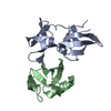
Mass: 18.015 Da / Num. of mol.: 226 / Source method: isolated from a natural source / Formula: H2O

-
Experimental details

-
Experiment

ExperimentMethod: X-RAY DIFFRACTION / Number of used crystals: 1

-
Sample preparation

CrystalDensity Matthews: 1.89 Å3/Da / Density % sol: 34.94 %
Crystal growTemperature: 296 K / Method: vapor diffusion, sitting drop
Details: 0.1 M MES pH6.6, 0.2 M Trimethylamine N-oxide dehydrate, 7% w/v PEG2000

-
Data collection

DiffractionMean temperature: 100 K / Serial crystal experiment: N
Diffraction sourceSource: SYNCHROTRON / Site: CLSI / Beamline: 08ID-1 / Wavelength: 0.95372 Å
DetectorType: DECTRIS EIGER X 9M / Detector: PIXEL / Date: Dec 8, 2022 / Details: TITANIUM SILICATE FLAT MIRROR WITH PT
RadiationMonochromator: DOUBLE CRYSTAL / MULTILAYER MONOCHROMATOR SI(111)
Protocol: SINGLE WAVELENGTH / Monochromatic (M) / Laue (L): M / Scattering type: x-ray
Radiation wavelengthWavelength: 0.95372 Å / Relative weight: 1
ReflectionResolution: 1.16→25.34 Å / Num. obs: 60078 / % possible obs: 99.8 % / Redundancy: 7 % / Biso Wilson estimate: 13.55 Å2 / CC1/2: 0.995 / Rpim(I) all: 0.03459 / Rrim(I) all: 0.0931 / Rsym value: 0.08622 / Net I/σ(I): 12
Reflection shellResolution: 1.16→1.18 Å / Mean I/σ(I) obs: 3.35 / Num. unique obs: 5857 / CC1/2: 0.873

-
Processing

Software
NameVersionClassification
REFMAC5.8.0352refinement
Cootmodel building
SCALAdata scaling
PHASERphasing
XDSdata reduction
RefinementMethod to determine structure: MOLECULAR REPLACEMENT / Resolution: 1.16→25.34 Å / Cross valid method: FREE R-VALUE
RfactorNum. reflection% reflection
Rfree0.2194 3013 5 %
Rwork0.1977 --
obs-60078 99.8 %
Displacement parametersBiso mean: 17.61 Å2
Refinement stepCycle: LAST / Resolution: 1.16→25.34 Å
ProteinNucleic acidLigandSolventTotal
Num. atoms1562 0 0 226 1788
LS refinement shellResolution: 1.16→1.18 Å
RfactorNum. reflection% reflection
Rfree0.235 --
Rwork0.229 --
obs-4115 99.1 %

+
About Yorodumi

-
News

-
Feb 9, 2022. New format data for meta-information of EMDB entries

New format data for meta-information of EMDB entries

  • Version 3 of the EMDB header file is now the official format.
  • The previous official version 1.9 will be removed from the archive.

Related info.:EMDB header

External links:wwPDB to switch to version 3 of the EMDB data model

-
Aug 12, 2020. Covid-19 info

Covid-19 info

URL: https://pdbjlvh1.pdbj.org/emnavi/covid19.php

New page: Covid-19 featured information page in EM Navigator.

Related info.:Covid-19 info / Mar 5, 2020. Novel coronavirus structure data

+
Mar 5, 2020. Novel coronavirus structure data

Novel coronavirus structure data

Related info.:Yorodumi Speices / Aug 12, 2020. Covid-19 info

External links:COVID-19 featured content - PDBj / Molecule of the Month (242):Coronavirus Proteases

+
Jan 31, 2019. EMDB accession codes are about to change! (news from PDBe EMDB page)

EMDB accession codes are about to change! (news from PDBe EMDB page)

  • The allocation of 4 digits for EMDB accession codes will soon come to an end. Whilst these codes will remain in use, new EMDB accession codes will include an additional digit and will expand incrementally as the available range of codes is exhausted. The current 4-digit format prefixed with “EMD-” (i.e. EMD-XXXX) will advance to a 5-digit format (i.e. EMD-XXXXX), and so on. It is currently estimated that the 4-digit codes will be depleted around Spring 2019, at which point the 5-digit format will come into force.
  • The EM Navigator/Yorodumi systems omit the EMD- prefix.

Related info.:Q: What is EMD? / ID/Accession-code notation in Yorodumi/EM Navigator

External links:EMDB Accession Codes are Changing Soon! / Contact to PDBj

+
Jul 12, 2017. Major update of PDB

Major update of PDB

  • wwPDB released updated PDB data conforming to the new PDBx/mmCIF dictionary.
  • This is a major update changing the version number from 4 to 5, and with Remediation, in which all the entries are updated.
  • In this update, many items about electron microscopy experimental information are reorganized (e.g. em_software).
  • Now, EM Navigator and Yorodumi are based on the updated data.

External links:wwPDB Remediation / Enriched Model Files Conforming to OneDep Data Standards Now Available in the PDB FTP Archive

-
Yorodumi

Thousand views of thousand structures

  • Yorodumi is a browser for structure data from EMDB, PDB, SASBDB, etc.
  • This page is also the successor to EM Navigator detail page, and also detail information page/front-end page for Omokage search.
  • The word "yorodu" (or yorozu) is an old Japanese word meaning "ten thousand". "mi" (miru) is to see.

Related info.:EMDB / PDB / SASBDB / Comparison of 3 databanks / Yorodumi Search / Aug 31, 2016. New EM Navigator & Yorodumi / Yorodumi Papers / Jmol/JSmol / Function and homology information / Changes in new EM Navigator and Yorodumi

Read more