 ムービー
ムービー コントローラー


+ データを開く

- 基本情報
基本情報
| 登録情報 | データベース: PDB / ID: 8ssk | ||||||||||||
|---|---|---|---|---|---|---|---|---|---|---|---|---|---|
| タイトル | Citrobacter rodentium contact dependent growth inhibition (CDI) entry and toxin (CdiA-CT) domains | ||||||||||||
 要素 要素 | Fimbrial usher protein StbD | ||||||||||||
 キーワード キーワード | TOXIN / Peptidase | ||||||||||||
| 機能・相同性 |  機能・相同性情報 機能・相同性情報 | ||||||||||||
| 生物種 |  Citrobacter rodentium (バクテリア) Citrobacter rodentium (バクテリア) | ||||||||||||
| 手法 |  X線回折 / シンクロトロン / 分子置換 / 解像度: 1.9 Å X線回折 / シンクロトロン / 分子置換 / 解像度: 1.9 Å | ||||||||||||
 データ登録者 データ登録者 | Cuthbert, B.J. / Goulding, C.W. / Hayes, C.S. / Nhan, D.Q. | ||||||||||||
| 資金援助 |  米国, 1件 米国, 1件
| ||||||||||||
 引用 引用 | ジャーナル: To Be Published タイトル: Citrobacter rodentium contact dependent growth inhibition (CDI) entry and toxin (CdiA-CT) domains 著者: Cuthbert, B.J. / Goulding, C.W. / Hayes, C.S. / Nhan, D.Q. | ||||||||||||
| 履歴 |
|
- 構造の表示
構造の表示
| 構造ビューア | 分子: MolmilJmol/JSmol |
|---|
- ダウンロードとリンク
-ダウンロード
| PDBx/mmCIF形式 | 8ssk.cif.gz | 556.9 KB | 表示 | PDBx/mmCIF形式 |
|---|---|---|---|---|
| PDB形式 | pdb8ssk.ent.gz | 368.7 KB | 表示 | PDB形式 |
| PDBx/mmJSON形式 | 8ssk.json.gz | ツリー表示 | PDBx/mmJSON形式 | |
| その他 |  その他のダウンロード その他のダウンロード |
-検証レポート
| アーカイブディレクトリ | https://data.pdbj.org/pub/pdb/validation_reports/ss/8sskftp://data.pdbj.org/pub/pdb/validation_reports/ss/8ssk | HTTPS FTP |
|---|
-関連構造データ

-リンク
 PDBj
PDBj

- 集合体
集合体
| 登録構造単位 | 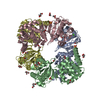
| ||||||||||||
|---|---|---|---|---|---|---|---|---|---|---|---|---|---|
| 1 | 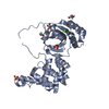
| ||||||||||||
| 2 | 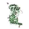
| ||||||||||||
| 3 | 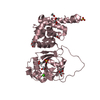
| ||||||||||||
| 4 | 
| ||||||||||||
| 5 |
| ||||||||||||
| 単位格子 |
|
-要素
-タンパク質 , 1種, 4分子 ABCD
| #1: タンパク質 | 分子量: 33576.453 Da / 分子数: 4 / 由来タイプ: 組換発現 由来: (組換発現) Citrobacter rodentium (バクテリア)遺伝子: stbD, E2R62_11320 / 発現宿主: |
|---|
-非ポリマー , 5種, 1283分子 


| #2: 化合物 | ChemComp-SO4 /  分子量: 96.063 Da / 分子数: 4 / 由来タイプ: 合成 / 式: SO4 分子量: 96.063 Da / 分子数: 4 / 由来タイプ: 合成 / 式: SO4#3: 化合物 | ChemComp-GOL /  分子量: 92.094 Da / 分子数: 11 / 由来タイプ: 合成 / 式: C3H8O3 分子量: 92.094 Da / 分子数: 11 / 由来タイプ: 合成 / 式: C3H8O3#4: 化合物 | ChemComp-ACT /  分子量: 59.044 Da / 分子数: 14 / 由来タイプ: 合成 / 式: C2H3O2 分子量: 59.044 Da / 分子数: 14 / 由来タイプ: 合成 / 式: C2H3O2#5: 化合物 | ChemComp-CA /  分子量: 40.078 Da / 分子数: 4 / 由来タイプ: 合成 / 式: Ca 分子量: 40.078 Da / 分子数: 4 / 由来タイプ: 合成 / 式: Ca#6: 水 | ChemComp-HOH / | 分子量: 18.015 Da / 分子数: 1250 / 由来タイプ: 天然 / 式: H2O |
|---|
-詳細
| 研究の焦点であるリガンドがあるか | N |
|---|---|
| Has protein modification | Y |
-実験情報
-実験
| 実験 | 手法: X線回折 / 使用した結晶の数: 1 |
|---|
- 試料調製
試料調製
| 結晶 | マシュー密度: 2.48 Å3/Da / 溶媒含有率: 50.33 % |
|---|---|
| 結晶化 | 温度: 277 K / 手法: 蒸気拡散法, ハンギングドロップ法 / pH: 4 / 詳細: 0.1 M sodium acetate, pH 4, 2 M ammonium sulfate |
-データ収集
| 回折 | 平均測定温度: 100 K / Serial crystal experiment: N |
|---|---|
| 放射光源 | 由来: シンクロトロン / サイト: SSRL / ビームライン: BL12-2 / 波長: 1 Å |
| 検出器 | タイプ: DECTRIS PILATUS 6M / 検出器: PIXEL / 日付: 2022年2月6日 |
| 放射 | プロトコル: SINGLE WAVELENGTH / 単色(M)・ラウエ(L): M / 散乱光タイプ: x-ray |
| 放射波長 | 波長: 1 Å / 相対比: 1 |
| 反射 | 解像度: 1.9→39.26 Å / Num. obs: 101283 / % possible obs: 98.5 % / 冗長度: 7 % / Biso Wilson estimate: 22.87 Å2 / CC1/2: 0.998 / Rmerge(I) obs: 0.118 / Rpim(I) all: 0.074 / Rrim(I) all: 0.14 / Χ2: 0.98 / Net I/σ(I): 11.4 |
| 反射 シェル | 解像度: 1.9→1.93 Å / 冗長度: 7 % / Rmerge(I) obs: 1.187 / Mean I/σ(I) obs: 1.6 / Num. unique obs: 4996 / CC1/2: 0.746 / Rpim(I) all: 0.748 / Rrim(I) all: 1.409 / Χ2: 0.99 / % possible all: 98.7 |
- 解析
解析
| ソフトウェア |
| |||||||||||||||||||||||||||||||||||||||||||||||||||||||||||||||||||||||||||||||||||||||||||||||||||||||||||||||||||||||||||||||||||||||||||||||||||||||||||||||||||||||||||||||||||||||||||||||||||||||||||||||||||||||||
|---|---|---|---|---|---|---|---|---|---|---|---|---|---|---|---|---|---|---|---|---|---|---|---|---|---|---|---|---|---|---|---|---|---|---|---|---|---|---|---|---|---|---|---|---|---|---|---|---|---|---|---|---|---|---|---|---|---|---|---|---|---|---|---|---|---|---|---|---|---|---|---|---|---|---|---|---|---|---|---|---|---|---|---|---|---|---|---|---|---|---|---|---|---|---|---|---|---|---|---|---|---|---|---|---|---|---|---|---|---|---|---|---|---|---|---|---|---|---|---|---|---|---|---|---|---|---|---|---|---|---|---|---|---|---|---|---|---|---|---|---|---|---|---|---|---|---|---|---|---|---|---|---|---|---|---|---|---|---|---|---|---|---|---|---|---|---|---|---|---|---|---|---|---|---|---|---|---|---|---|---|---|---|---|---|---|---|---|---|---|---|---|---|---|---|---|---|---|---|---|---|---|---|---|---|---|---|---|---|---|---|---|---|---|---|---|---|---|---|
| 精密化 | 構造決定の手法: 分子置換 / 解像度: 1.9→39.26 Å / SU ML: 0.1846 / 交差検証法: FREE R-VALUE / σ(F): 1.35 / 位相誤差: 19.245 立体化学のターゲット値: GeoStd + Monomer Library + CDL v1.2
| |||||||||||||||||||||||||||||||||||||||||||||||||||||||||||||||||||||||||||||||||||||||||||||||||||||||||||||||||||||||||||||||||||||||||||||||||||||||||||||||||||||||||||||||||||||||||||||||||||||||||||||||||||||||||
| 溶媒の処理 | 減衰半径: 0.9 Å / VDWプローブ半径: 1.1 Å / 溶媒モデル: FLAT BULK SOLVENT MODEL | |||||||||||||||||||||||||||||||||||||||||||||||||||||||||||||||||||||||||||||||||||||||||||||||||||||||||||||||||||||||||||||||||||||||||||||||||||||||||||||||||||||||||||||||||||||||||||||||||||||||||||||||||||||||||
| 原子変位パラメータ | Biso mean: 28.67 Å2 | |||||||||||||||||||||||||||||||||||||||||||||||||||||||||||||||||||||||||||||||||||||||||||||||||||||||||||||||||||||||||||||||||||||||||||||||||||||||||||||||||||||||||||||||||||||||||||||||||||||||||||||||||||||||||
| 精密化ステップ | サイクル: LAST / 解像度: 1.9→39.26 Å
| |||||||||||||||||||||||||||||||||||||||||||||||||||||||||||||||||||||||||||||||||||||||||||||||||||||||||||||||||||||||||||||||||||||||||||||||||||||||||||||||||||||||||||||||||||||||||||||||||||||||||||||||||||||||||
| 拘束条件 |
| |||||||||||||||||||||||||||||||||||||||||||||||||||||||||||||||||||||||||||||||||||||||||||||||||||||||||||||||||||||||||||||||||||||||||||||||||||||||||||||||||||||||||||||||||||||||||||||||||||||||||||||||||||||||||
| LS精密化 シェル |
|
