Entry Database : PDB / ID : 8ssdTitle Methionine synthase, C-terminal fragment, Cobalamin and Reactivation Domains from Thermus thermophilus HB8 Methionine synthase Keywords / / / / / Function / homology Function Domain/homology Component
/ / / / / / / / / / / / / / / / / / / / / / / / / / / / / / / / / / / / / / / Biological species Thermus thermophilus HB8 (bacteria)Method / / / Resolution : 2.4 Å Authors Yamada, K. / Mendoza, J. / Koutmos, M. Funding support Organization Grant number Country National Science Foundation (NSF, United States) 1945174
Journal : Nat Commun / Year : 2023Title : Structure of full-length cobalamin-dependent methionine synthase and cofactor loading captured in crystallo.Authors : Mendoza, J. / Purchal, M. / Yamada, K. / Koutmos, M. History Deposition May 8, 2023 Deposition site / Processing site Revision 1.0 Oct 11, 2023 Provider / Type Revision 1.1 Oct 18, 2023 Group / Category Item _citation.country / _citation.journal_abbrev ... _citation.country / _citation.journal_abbrev / _citation.journal_id_ISSN / _citation.journal_volume / _citation.page_first / _citation.pdbx_database_id_DOI / _citation.title Revision 1.2 May 1, 2024 Group / Category Item / _citation.pdbx_database_id_PubMed / _citation.title
Show all Show less
 Movie
Movie Controller

 Yorodumi
 Open data
 Basic information
 Components
 Keywords
 Function and homology information
 Thermus thermophilus HB8 (bacteria)
 X-RAY DIFFRACTION /
  Authors
 United States, 1items
  Citation
 Structure visualization
 Downloads & links
 Other downloads
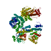


 PDBj
PDBj Assembly
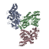
 Mass: 18.015 Da / Num. of mol.: 485 / Source method: isolated from a natural source / Formula: H2O
 Sample preparation
 Processing