 Movie
Movie Controller

[English] 日本語
 Yorodumi
- PDB-8srt: Crystal structure of the O-acetyl-L-serine sulfhydrylase A (CysK)... -
+ Open data

- Basic information
| Entry | Database: PDB / ID: 8srt | ||||||
|---|---|---|---|---|---|---|---|
| Title | Crystal structure of the O-acetyl-L-serine sulfhydrylase A (CysK) holoenzyme from Staphylococcus aureus NCTC 8325 | ||||||
 Components Components | Cysteine synthase | ||||||
 Keywords Keywords | TRANSFERASE / Pyridoxal-phosphate dependent enzyme | ||||||
| Function / homology |  Function and homology information Function and homology informationL-cysteine desulfhydrase activity / cysteine synthase / cysteine synthase activity / L-cysteine biosynthetic process from L-serine / cytoplasm Similarity search - Function | ||||||
| Biological species |  Staphylococcus aureus subsp. aureus NCTC 8325 (bacteria) Staphylococcus aureus subsp. aureus NCTC 8325 (bacteria) | ||||||
| Method |  X-RAY DIFFRACTION / SYNCHROTRON / MOLECULAR REPLACEMENT / molecular replacement / Resolution: 1.9 Å X-RAY DIFFRACTION / SYNCHROTRON / MOLECULAR REPLACEMENT / molecular replacement / Resolution: 1.9 Å | ||||||
 Authors Authors | Pederick, J.L. / Vandborg, B.C. / Bruning, J.B. | ||||||
| Funding support | 1items
| ||||||
 Citation Citation | Journal: Acs Infect Dis. / Year: 2025 Title: Identification of Cysteine Metabolism Regulator (CymR)-Derived Pentapeptides as Nanomolar Inhibitors of Staphylococcus aureus O -Acetyl-l-serine Sulfhydrylase (CysK). Authors: Pederick, J.L. / Vandborg, B.C. / George, A. / Bovermann, H. / Boyd, J.M. / Freundlich, J.S. / Bruning, J.B. | ||||||
| History |
|
- Structure visualization
| Structure viewer | Molecule: MolmilJmol/JSmol |
|---|
- Downloads & links
-Download
| PDBx/mmCIF format | 8srt.cif.gz | 131.2 KB | Display | PDBx/mmCIF format |
|---|---|---|---|---|
| PDB format | pdb8srt.ent.gz | 98.2 KB | Display | PDB format |
| PDBx/mmJSON format | 8srt.json.gz | Tree view | PDBx/mmJSON format | |
| Others |  Other downloads Other downloads |
-Validation report
| Summary document | 8srt_validation.pdf.gz | 430.5 KB | Display | wwPDB validaton report |
|---|---|---|---|---|
| Full document | 8srt_full_validation.pdf.gz | 435.3 KB | Display | |
| Data in XML | 8srt_validation.xml.gz | 29.3 KB | Display | |
| Data in CIF | 8srt_validation.cif.gz | 39.1 KB | Display | |
| Arichive directory | https://data.pdbj.org/pub/pdb/validation_reports/sr/8srtftp://data.pdbj.org/pub/pdb/validation_reports/sr/8srt | HTTPS FTP |
-Related structure data
| Related structure data |  8sruC  8srvC  8srwC  8t2cC 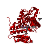 2q3bS S: Starting model for refinement C: citing same article ( |
|---|---|
| Similar structure data |
-Links
 PDBj
PDBj
- Assembly
| Deposited unit | 
| ||||||||
|---|---|---|---|---|---|---|---|---|---|
| 1 |
| ||||||||
| Unit cell |
|
-Components
| #1: Protein | Mass: 34257.852 Da / Num. of mol.: 2 Source method: isolated from a genetically manipulated source Source: (gene. exp.) Staphylococcus aureus subsp. aureus NCTC 8325 (bacteria)Gene: SAOUHSC_00488 / Production host: #2: Water | ChemComp-HOH / |  Mass: 18.015 Da / Num. of mol.: 233 / Source method: isolated from a natural source / Formula: H2O Mass: 18.015 Da / Num. of mol.: 233 / Source method: isolated from a natural source / Formula: H2OHas ligand of interest | N | Has protein modification | Y | |
|---|
-Experimental details
-Experiment
| Experiment | Method: X-RAY DIFFRACTION / Number of used crystals: 1 |
|---|
- Sample preparation
| Crystal | Density Matthews: 2.09 Å3/Da / Density % sol: 41.12 % |
|---|---|
| Crystal grow | Temperature: 289.15 K / Method: vapor diffusion, sitting drop Details: 0.2 M ammonium acetate, 0.1 M HEPES pH 7.5, 25% PEG 3350 |
-Data collection
| Diffraction | Mean temperature: 100 K / Serial crystal experiment: N | ||||||||||||||||||||||||||||||
|---|---|---|---|---|---|---|---|---|---|---|---|---|---|---|---|---|---|---|---|---|---|---|---|---|---|---|---|---|---|---|---|
| Diffraction source | Source: SYNCHROTRON / Site: Australian Synchrotron  / Beamline: MX1 / Wavelength: 0.9537 Å / Beamline: MX1 / Wavelength: 0.9537 Å | ||||||||||||||||||||||||||||||
| Detector | Type: DECTRIS EIGER2 X 9M / Detector: PIXEL / Date: Feb 17, 2021 | ||||||||||||||||||||||||||||||
| Radiation | Protocol: SINGLE WAVELENGTH / Monochromatic (M) / Laue (L): M / Scattering type: x-ray | ||||||||||||||||||||||||||||||
| Radiation wavelength | Wavelength: 0.9537 Å / Relative weight: 1 | ||||||||||||||||||||||||||||||
| Reflection | Resolution: 1.9→35.95 Å / Num. obs: 44294 / % possible obs: 99.8 % / Redundancy: 6.8 % / CC1/2: 0.999 / Rmerge(I) obs: 0.063 / Rpim(I) all: 0.026 / Rrim(I) all: 0.069 / Net I/σ(I): 13 / Num. measured all: 301349 / Scaling rejects: 54 | ||||||||||||||||||||||||||||||
| Reflection shell | Diffraction-ID: 1
|
-Phasing
| Phasing | Method: molecular replacement |
|---|
- Processing
| Software |
| |||||||||||||||||||||||||||||||||||||||||||||||||||||||||||||||||||||||||||||||||||||||||||||||||||||||||||||||||||||||
|---|---|---|---|---|---|---|---|---|---|---|---|---|---|---|---|---|---|---|---|---|---|---|---|---|---|---|---|---|---|---|---|---|---|---|---|---|---|---|---|---|---|---|---|---|---|---|---|---|---|---|---|---|---|---|---|---|---|---|---|---|---|---|---|---|---|---|---|---|---|---|---|---|---|---|---|---|---|---|---|---|---|---|---|---|---|---|---|---|---|---|---|---|---|---|---|---|---|---|---|---|---|---|---|---|---|---|---|---|---|---|---|---|---|---|---|---|---|---|---|---|
| Refinement | Method to determine structure: MOLECULAR REPLACEMENT Starting model: 2Q3B Resolution: 1.9→35.95 Å / SU ML: 0.22 / Cross valid method: THROUGHOUT / σ(F): 1.34 / Phase error: 25.83 / Stereochemistry target values: ML
| |||||||||||||||||||||||||||||||||||||||||||||||||||||||||||||||||||||||||||||||||||||||||||||||||||||||||||||||||||||||
| Solvent computation | Shrinkage radii: 0.9 Å / VDW probe radii: 1.11 Å / Solvent model: FLAT BULK SOLVENT MODEL | |||||||||||||||||||||||||||||||||||||||||||||||||||||||||||||||||||||||||||||||||||||||||||||||||||||||||||||||||||||||
| Displacement parameters | Biso max: 94 Å2 / Biso mean: 43.7348 Å2 / Biso min: 25.58 Å2 | |||||||||||||||||||||||||||||||||||||||||||||||||||||||||||||||||||||||||||||||||||||||||||||||||||||||||||||||||||||||
| Refinement step | Cycle: final / Resolution: 1.9→35.95 Å
| |||||||||||||||||||||||||||||||||||||||||||||||||||||||||||||||||||||||||||||||||||||||||||||||||||||||||||||||||||||||
| LS refinement shell | Refine-ID: X-RAY DIFFRACTION / Rfactor Rfree error: 0 / Total num. of bins used: 16
|
