 Movie
Movie Controller

[English] 日本語
 Yorodumi
- PDB-8sjx: Structure of lens aquaporin-0 array in sphingomyelin/cholesterol ... -
+ Open data

- Basic information
| Entry | Database: PDB / ID: 8sjx | ||||||
|---|---|---|---|---|---|---|---|
| Title | Structure of lens aquaporin-0 array in sphingomyelin/cholesterol bilayer (2SM:1Chol) | ||||||
 Components Components | Lens fiber major intrinsic protein | ||||||
 Keywords Keywords | MEMBRANE PROTEIN / aquaporin / lens / cholesterol / lipid raft | ||||||
| Function / homology |  Function and homology information Function and homology informationmaintenance of lens transparency / homotypic cell-cell adhesion / gap junction-mediated intercellular transport / water channel activity / cell adhesion mediator activity / structural constituent of eye lens / water transport / anchoring junction / lens development in camera-type eye / visual perception ...maintenance of lens transparency / homotypic cell-cell adhesion / gap junction-mediated intercellular transport / water channel activity / cell adhesion mediator activity / structural constituent of eye lens / water transport / anchoring junction / lens development in camera-type eye / visual perception / calmodulin binding / apical plasma membrane / plasma membrane Similarity search - Function | ||||||
| Biological species |  | ||||||
| Method | ELECTRON CRYSTALLOGRAPHY / electron crystallography /  MOLECULAR REPLACEMENT / Resolution: 2.5 Å MOLECULAR REPLACEMENT / Resolution: 2.5 Å | ||||||
 Authors Authors | Chiu, P.-L. / Walz, T. | ||||||
| Funding support |  United States, 1items United States, 1items
| ||||||
 Citation Citation | Journal: Elife / Year: 2024 Title: Structure and dynamics of cholesterol-mediated aquaporin-0 arrays and implications for lipid rafts. Authors: Po-Lin Chiu / Juan D Orjuela / Bert L de Groot / Camilo Aponte Santamaría / Thomas Walz /  Abstract: Aquaporin-0 (AQP0) tetramers form square arrays in lens membranes through a yet unknown mechanism, but lens membranes are enriched in sphingomyelin and cholesterol. Here, we determined electron ...Aquaporin-0 (AQP0) tetramers form square arrays in lens membranes through a yet unknown mechanism, but lens membranes are enriched in sphingomyelin and cholesterol. Here, we determined electron crystallographic structures of AQP0 in sphingomyelin/cholesterol membranes and performed molecular dynamics (MD) simulations to establish that the observed cholesterol positions represent those seen around an isolated AQP0 tetramer and that the AQP0 tetramer largely defines the location and orientation of most of its associated cholesterol molecules. At a high concentration, cholesterol increases the hydrophobic thickness of the annular lipid shell around AQP0 tetramers, which may thus cluster to mitigate the resulting hydrophobic mismatch. Moreover, neighboring AQP0 tetramers sandwich a cholesterol deep in the center of the membrane. MD simulations show that the association of two AQP0 tetramers is necessary to maintain the deep cholesterol in its position and that the deep cholesterol increases the force required to laterally detach two AQP0 tetramers, not only due to protein-protein contacts but also due to increased lipid-protein complementarity. Since each tetramer interacts with four such 'glue' cholesterols, avidity effects may stabilize larger arrays. The principles proposed to drive AQP0 array formation could also underlie protein clustering in lipid rafts. #1: Journal: Elife / Year: 2024Title: Structure and dynamics of cholesterol-mediated aquaporin-0 arrays and implications for lipid rafts Authors: Chiu, P.L. / Orjuela, J.D. / de Groot, B.L. / Aponte-Santamaria, C. / Walz, T. | ||||||
| History |
|
- Structure visualization
| Structure viewer | Molecule: MolmilJmol/JSmol |
|---|
- Downloads & links
-Download
| PDBx/mmCIF format | 8sjx.cif.gz | 78.6 KB | Display | PDBx/mmCIF format |
|---|---|---|---|---|
| PDB format | pdb8sjx.ent.gz | 47.5 KB | Display | PDB format |
| PDBx/mmJSON format | 8sjx.json.gz | Tree view | PDBx/mmJSON format | |
| Others |  Other downloads Other downloads |
-Validation report
| Arichive directory | https://data.pdbj.org/pub/pdb/validation_reports/sj/8sjxftp://data.pdbj.org/pub/pdb/validation_reports/sj/8sjx | HTTPS FTP |
|---|
-Related structure data

-Links
 PDBj
PDBj


- Assembly
| Deposited unit | 
| ||||||||||
|---|---|---|---|---|---|---|---|---|---|---|---|
| 1 | x 8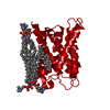
| ||||||||||
| Unit cell |
|
-Components
| #1: Protein | Mass: 28284.885 Da / Num. of mol.: 1 / Source method: isolated from a natural source / Source: (natural) | ||||||||
|---|---|---|---|---|---|---|---|---|---|
| #2: Chemical | ChemComp-HWP / [(   Mass: 703.028 Da / Num. of mol.: 7 / Source method: obtained synthetically / Formula: C39H79N2O6P / Feature type: SUBJECT OF INVESTIGATION Mass: 703.028 Da / Num. of mol.: 7 / Source method: obtained synthetically / Formula: C39H79N2O6P / Feature type: SUBJECT OF INVESTIGATION#3: Chemical | ChemComp-CLR / |   Mass: 386.654 Da / Num. of mol.: 1 / Source method: obtained synthetically / Formula: C27H46O / Feature type: SUBJECT OF INVESTIGATION Mass: 386.654 Da / Num. of mol.: 1 / Source method: obtained synthetically / Formula: C27H46O / Feature type: SUBJECT OF INVESTIGATION#4: Water | ChemComp-HOH / |  Mass: 18.015 Da / Num. of mol.: 11 / Source method: isolated from a natural source / Formula: H2O Mass: 18.015 Da / Num. of mol.: 11 / Source method: isolated from a natural source / Formula: H2OHas ligand of interest | Y | Has protein modification | N | |
-Experimental details
-Experiment
| Experiment | Method: ELECTRON CRYSTALLOGRAPHY |
|---|---|
| EM experiment | Aggregation state: 2D ARRAY / 3D reconstruction method: electron crystallography |
| Crystal symmetry | ∠γ: 90 ° / C sampling length: 200 Å / A: 65.5 Å / B: 65.5 Å / C: 200 Å / Space group name H-M: P422 |
- Sample preparation
| Component | Name: Lens aquaporin-0 in sphingomyelin/cholesterol bilayer / Type: COMPLEX / Entity ID: #1 / Source: NATURAL | |||||||||||||||||||||||||
|---|---|---|---|---|---|---|---|---|---|---|---|---|---|---|---|---|---|---|---|---|---|---|---|---|---|---|
| Molecular weight | Value: 0.0283 MDa / Experimental value: NO | |||||||||||||||||||||||||
| Source (natural) | Organism: | |||||||||||||||||||||||||
| EM crystal formation | Lipid mixture: Sphingomyelin and cholesterol were mixed at a 2:1 molar ratio. Lipid protein ratio: 0.2 / Temperature: 310 K / Time: 7 DAY | |||||||||||||||||||||||||
| Buffer solution | pH: 6 | |||||||||||||||||||||||||
| Buffer component |
| |||||||||||||||||||||||||
| Specimen | Embedding applied: YES / Shadowing applied: NO / Staining applied: NO / Vitrification applied: NO / Details: 2D crystal of lens AQP0. | |||||||||||||||||||||||||
| Specimen support | Grid material: MOLYBDENUM / Grid type: Homemade | |||||||||||||||||||||||||
| EM embedding | Details: Aquaporin-0 2D crystals were prepared on molybdenum grids using the carbon sandwich method and a trehalose concentration ranging from 3% to 5% (w/v). Material: Trehalose |
-Data collection
| Experimental equipment |  Model: Tecnai Polara / Image courtesy: FEI Company |
|---|---|
| Microscopy | Model: FEI POLARA 300 Details: The diffraction patterns were recorded without setting defocus. |
| Electron gun | Electron source: FIELD EMISSION GUN / Accelerating voltage: 300 kV / Illumination mode: FLOOD BEAM |
| Electron lens | Mode: DIFFRACTION / Nominal defocus max: 0 nm / Nominal defocus min: 0 nm / Calibrated defocus min: 0 nm / Calibrated defocus max: 0 nm / C2 aperture diameter: 30 µm / Alignment procedure: COMA FREE |
| Specimen holder | Cryogen: NITROGEN / Specimen holder model: OTHER |
| Image recording | Average exposure time: 30 sec. / Electron dose: 10 e/Å2 / Film or detector model: GATAN ULTRASCAN 4000 (4k x 4k) / Num. of diffraction images: 214 |
| EM diffraction shell | Resolution: 2.3→13.8 Å / Fourier space coverage: 86.71 % / Multiplicity: 6.2 / Num. of structure factors: 16437 / Phase residual: 1.0E-5 ° |
| EM diffraction stats | Details: There was no phase error rejection criteria used for diffraction intensities. Fourier space coverage: 86.71 % / High resolution: 2.3 Å / Num. of intensities measured: 122501 / Num. of structure factors: 16437 / Phase error rejection criteria: 0 / Rmerge: 21.6 / Rsym: 14.9 |
| Reflection | Biso Wilson estimate: 49.13 Å2 |
- Processing
| Software |
| |||||||||||||||||||||||||||||||||||||||||||||||||||||||||||||||||||||||||||||||||||||||||||
|---|---|---|---|---|---|---|---|---|---|---|---|---|---|---|---|---|---|---|---|---|---|---|---|---|---|---|---|---|---|---|---|---|---|---|---|---|---|---|---|---|---|---|---|---|---|---|---|---|---|---|---|---|---|---|---|---|---|---|---|---|---|---|---|---|---|---|---|---|---|---|---|---|---|---|---|---|---|---|---|---|---|---|---|---|---|---|---|---|---|---|---|---|
| EM software |
| |||||||||||||||||||||||||||||||||||||||||||||||||||||||||||||||||||||||||||||||||||||||||||
| Crystal symmetry | ∠γ: 90 ° / C sampling length: 200 Å / A: 65.5 Å / B: 65.5 Å / C: 200 Å / Space group name H-M: P422 | |||||||||||||||||||||||||||||||||||||||||||||||||||||||||||||||||||||||||||||||||||||||||||
| CTF correction | Type: NONE | |||||||||||||||||||||||||||||||||||||||||||||||||||||||||||||||||||||||||||||||||||||||||||
| 3D reconstruction | Resolution: 2.5 Å / Resolution method: DIFFRACTION PATTERN/LAYERLINES / Symmetry type: 2D CRYSTAL | |||||||||||||||||||||||||||||||||||||||||||||||||||||||||||||||||||||||||||||||||||||||||||
| Atomic model building | PDB-ID: 2B6O Pdb chain-ID: A / Accession code: 2B6O / Chain residue range: 6-255 / Details: Molecular replacement / Pdb chain residue range: 6-255 / Source name: PDB / Type: experimental model | |||||||||||||||||||||||||||||||||||||||||||||||||||||||||||||||||||||||||||||||||||||||||||
| Refinement | Method to determine structure: MOLECULAR REPLACEMENT / Resolution: 2.5→2.5 Å / SU ML: 0.3971 / Cross valid method: FREE R-VALUE / σ(F): 1.33 / Phase error: 33.7767 Stereochemistry target values: GeoStd + Monomer Library + CDL v1.2
| |||||||||||||||||||||||||||||||||||||||||||||||||||||||||||||||||||||||||||||||||||||||||||
| Solvent computation | Shrinkage radii: 0.9 Å / VDW probe radii: 1.1 Å / Solvent model: FLAT BULK SOLVENT MODEL | |||||||||||||||||||||||||||||||||||||||||||||||||||||||||||||||||||||||||||||||||||||||||||
| Displacement parameters | Biso mean: 66.6 Å2 | |||||||||||||||||||||||||||||||||||||||||||||||||||||||||||||||||||||||||||||||||||||||||||
| Refine LS restraints |
| |||||||||||||||||||||||||||||||||||||||||||||||||||||||||||||||||||||||||||||||||||||||||||
| LS refinement shell |
|

