 Movie
Movie Controller


+ Open data

- Basic information
| Entry | Database: PDB / ID: 8sjk | ||||||
|---|---|---|---|---|---|---|---|
| Title | Pembrolizumab Caffeine crystal | ||||||
 Components Components |
| ||||||
 Keywords Keywords | IMMUNE SYSTEM / CANCER / ANTIBODY / SUBCLASSES / IGG4 / MELANOMA / NSCLC | ||||||
| Function / homology | sucrose / CAFFEINE Function and homology information Function and homology information | ||||||
| Biological species |  Homo sapiens (human) Homo sapiens (human) | ||||||
| Method |  X-RAY DIFFRACTION / SYNCHROTRON / MOLECULAR REPLACEMENT / Resolution: 2.22 Å X-RAY DIFFRACTION / SYNCHROTRON / MOLECULAR REPLACEMENT / Resolution: 2.22 Å | ||||||
 Authors Authors | Larpent, P. / Codan, L. / Bothe, J.R. / Stueber, D. / Reichert, P. / Fischmann, T. / Su, Y. / Pabit, S. / Gupta, S. / Iuzzolino, L. / Cote, A. | ||||||
| Funding support |  United States, 1items United States, 1items
| ||||||
 Citation Citation | Journal: Mol Pharm. / Year: 2024 Title: Small-Angle X-ray Scattering as a Powerful Tool for Phase and Crystallinity Assessment of Monoclonal Antibody Crystallites in Support of Batch Crystallization. Authors: Larpent, P. / Codan, L. / Bothe, J.R. / Iuzzolino, L. / Pabit, S. / Gupta, S. / Fischmann, T. / Su, Y. / Reichert, P. / Stueber, D. / Cote, A. | ||||||
| History |
|
- Structure visualization
| Structure viewer | Molecule: MolmilJmol/JSmol |
|---|
- Downloads & links
-Download
| PDBx/mmCIF format | 8sjk.cif.gz | 276.5 KB | Display | PDBx/mmCIF format |
|---|---|---|---|---|
| PDB format | pdb8sjk.ent.gz | 220.8 KB | Display | PDB format |
| PDBx/mmJSON format | 8sjk.json.gz | Tree view | PDBx/mmJSON format | |
| Others |  Other downloads Other downloads |
-Validation report
| Arichive directory | https://data.pdbj.org/pub/pdb/validation_reports/sj/8sjkftp://data.pdbj.org/pub/pdb/validation_reports/sj/8sjk | HTTPS FTP |
|---|
-Related structure data
| Similar structure data |
|---|
-Links
 PDBj
PDBj


- Assembly
| Deposited unit | 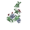
| ||||||||
|---|---|---|---|---|---|---|---|---|---|
| 1 | 
| ||||||||
| Unit cell |
|
-Components
-Antibody , 2 types, 2 molecules AB
| #1: Antibody | Mass: 23768.449 Da / Num. of mol.: 1 Source method: isolated from a genetically manipulated source Source: (gene. exp.) Homo sapiens (human) / Production host: Mammalia (mammals) |
|---|---|
| #2: Antibody | Mass: 49166.047 Da / Num. of mol.: 1 Source method: isolated from a genetically manipulated source Source: (gene. exp.) Homo sapiens (human) / Production host: Mammalia (mammals) |
-Sugars , 2 types, 3 molecules
| #3: Polysaccharide | 2-acetamido-2-deoxy-beta-D-glucopyranose-(1-2)-alpha-D-mannopyranose-(1-3)-[2-acetamido-2-deoxy- ...2-acetamido-2-deoxy-beta-D-glucopyranose-(1-2)-alpha-D-mannopyranose-(1-3)-[2-acetamido-2-deoxy-beta-D-glucopyranose-(1-2)-alpha-D-mannopyranose-(1-6)]beta-D-mannopyranose-(1-4)-2-acetamido-2-deoxy-beta-D-glucopyranose-(1-4)-2-acetamido-2-deoxy-beta-D-glucopyranose Source method: isolated from a genetically manipulated source |
|---|---|
| #4: Polysaccharide |   Source method: isolated from a genetically manipulated source Details: oligosaccharide with reducing-end-to-reducing-end glycosidic bond References: sucrose |
-Non-polymers , 2 types, 301 molecules 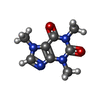

| #5: Chemical | ChemComp-CFF /  Mass: 194.191 Da / Num. of mol.: 1 / Source method: obtained synthetically / Formula: C8H10N4O2 / Feature type: SUBJECT OF INVESTIGATION / Comment: medication*YM Mass: 194.191 Da / Num. of mol.: 1 / Source method: obtained synthetically / Formula: C8H10N4O2 / Feature type: SUBJECT OF INVESTIGATION / Comment: medication*YM |
|---|---|
| #6: Water | ChemComp-HOH / Mass: 18.015 Da / Num. of mol.: 300 / Source method: isolated from a natural source / Formula: H2O |
-Details
| Has ligand of interest | Y |
|---|---|
| Has protein modification | Y |
-Experimental details
-Experiment
| Experiment | Method: X-RAY DIFFRACTION / Number of used crystals: 1 |
|---|
- Sample preparation
| Crystal | Density Matthews: 2.99 Å3/Da / Density % sol: 58.9 % |
|---|---|
| Crystal grow | Temperature: 300 K / Method: vapor diffusion / pH: 6.8 Details: 0.1M Na HEPES pH 6.8 0.2%W/V Caffeine, 12% PEG3350, 3% DEXTRAN |
-Data collection
| Diffraction | Mean temperature: 100 K / Serial crystal experiment: N |
|---|---|
| Diffraction source | Source: SYNCHROTRON / Site: APS / Beamline: 22-ID / Wavelength: 1 Å |
| Detector | Type: DECTRIS PILATUS 6M / Detector: PIXEL / Date: Jul 1, 2017 |
| Radiation | Protocol: SINGLE WAVELENGTH / Monochromatic (M) / Laue (L): M / Scattering type: x-ray |
| Radiation wavelength | Wavelength: 1 Å / Relative weight: 1 |
| Reflection | Resolution: 2.22→40.81 Å / Num. obs: 33863 / % possible obs: 99.7 % / Redundancy: 8.7 % / CC1/2: 0.995 / Rmerge(I) obs: 0.138 / Rrim(I) all: 0.147 / Net I/σ(I): 14.1 |
| Reflection shell | Resolution: 2.22→2.453 Å / Redundancy: 3.7 % / Rmerge(I) obs: 0.904 / Mean I/σ(I) obs: 1.3 / Num. unique obs: 2412 / CC1/2: 0.542 / Rpim(I) all: 0.048 / Rrim(I) all: 0.147 / % possible all: 15.1 |
- Processing
| Software |
| ||||||||||||||||||||||||||||||||||||||||||||||||||||||||||||||||||||||||||||||||||||||||||||||||||||||||||||||||||
|---|---|---|---|---|---|---|---|---|---|---|---|---|---|---|---|---|---|---|---|---|---|---|---|---|---|---|---|---|---|---|---|---|---|---|---|---|---|---|---|---|---|---|---|---|---|---|---|---|---|---|---|---|---|---|---|---|---|---|---|---|---|---|---|---|---|---|---|---|---|---|---|---|---|---|---|---|---|---|---|---|---|---|---|---|---|---|---|---|---|---|---|---|---|---|---|---|---|---|---|---|---|---|---|---|---|---|---|---|---|---|---|---|---|---|---|
| Refinement | Method to determine structure: MOLECULAR REPLACEMENT / Resolution: 2.22→40.81 Å / Cor.coef. Fo:Fc: 0.932 / Cor.coef. Fo:Fc free: 0.899 / Rfactor Rfree error: 0 / Cross valid method: THROUGHOUT / σ(F): 0 / SU R Blow DPI: 0.323 / SU Rfree Blow DPI: 0.234
| ||||||||||||||||||||||||||||||||||||||||||||||||||||||||||||||||||||||||||||||||||||||||||||||||||||||||||||||||||
| Displacement parameters | Biso mean: 37.21 Å2
| ||||||||||||||||||||||||||||||||||||||||||||||||||||||||||||||||||||||||||||||||||||||||||||||||||||||||||||||||||
| Refine analyze | Luzzati coordinate error obs: 0.29 Å | ||||||||||||||||||||||||||||||||||||||||||||||||||||||||||||||||||||||||||||||||||||||||||||||||||||||||||||||||||
| Refinement step | Cycle: 1 / Resolution: 2.22→40.81 Å
| ||||||||||||||||||||||||||||||||||||||||||||||||||||||||||||||||||||||||||||||||||||||||||||||||||||||||||||||||||
| Refine LS restraints |
| ||||||||||||||||||||||||||||||||||||||||||||||||||||||||||||||||||||||||||||||||||||||||||||||||||||||||||||||||||
| LS refinement shell | Resolution: 2.22→2.29 Å / Rfactor Rfree error: 0 / Total num. of bins used: 17
| ||||||||||||||||||||||||||||||||||||||||||||||||||||||||||||||||||||||||||||||||||||||||||||||||||||||||||||||||||
| Refinement TLS params. | Method: refined / Refine-ID: X-RAY DIFFRACTION
| ||||||||||||||||||||||||||||||||||||||||||||||||||||||||||||||||||||||||||||||||||||||||||||||||||||||||||||||||||
| Refinement TLS group |
|
