 Movie
Movie Controller

[English] 日本語
 Yorodumi
- PDB-8sjj: X-ray structure of the metastable SEPT14-SEPT7 heterodimeric coil... -
+ Open data

- Basic information
| Entry | Database: PDB / ID: 8sjj | |||||||||
|---|---|---|---|---|---|---|---|---|---|---|
| Title | X-ray structure of the metastable SEPT14-SEPT7 heterodimeric coiled coil | |||||||||
 Components Components |
| |||||||||
 Keywords Keywords | CELL CYCLE / Septin / Coiled coil | |||||||||
| Function / homology |  Function and homology information Function and homology informationseptin complex / protein localization to perinuclear region of cytoplasm / septin ring / cytoskeleton-dependent cytokinesis / motile cilium / cell division site / cilium assembly / spermatid development / acrosomal vesicle / kinetochore ...septin complex / protein localization to perinuclear region of cytoplasm / septin ring / cytoskeleton-dependent cytokinesis / motile cilium / cell division site / cilium assembly / spermatid development / acrosomal vesicle / kinetochore / neuron migration / intracellular protein localization / microtubule cytoskeleton / spermatogenesis / perikaryon / molecular adaptor activity / cytoskeleton / axon / GTPase activity / dendrite / GTP binding / perinuclear region of cytoplasm / cytoplasm Similarity search - Function | |||||||||
| Biological species |  Homo sapiens (human) Homo sapiens (human) | |||||||||
| Method |  X-RAY DIFFRACTION / SYNCHROTRON / MOLECULAR REPLACEMENT / Resolution: 1.781 Å X-RAY DIFFRACTION / SYNCHROTRON / MOLECULAR REPLACEMENT / Resolution: 1.781 Å | |||||||||
 Authors Authors | Cavini, I.A. / Pereira, H.M. / Garratt, R.C. | |||||||||
| Funding support |  Brazil, 2items Brazil, 2items
| |||||||||
 Citation Citation | Journal: Acta Crystallogr D Struct Biol / Year: 2023 Title: X-ray structure of the metastable SEPT14-SEPT7 coiled coil reveals a hendecad region crucial for heterodimerization. Authors: Cavini, I.A. / Winter, A.J. / D'Muniz Pereira, H. / Woolfson, D.N. / Crump, M.P. / Garratt, R.C. | |||||||||
| History |
|
- Structure visualization
| Structure viewer | Molecule: MolmilJmol/JSmol |
|---|
- Downloads & links
-Download
| PDBx/mmCIF format | 8sjj.cif.gz | 152.5 KB | Display | PDBx/mmCIF format |
|---|---|---|---|---|
| PDB format | pdb8sjj.ent.gz | 118.7 KB | Display | PDB format |
| PDBx/mmJSON format | 8sjj.json.gz | Tree view | PDBx/mmJSON format | |
| Others |  Other downloads Other downloads |
-Validation report
| Summary document | 8sjj_validation.pdf.gz | 3 MB | Display | wwPDB validaton report |
|---|---|---|---|---|
| Full document | 8sjj_full_validation.pdf.gz | 3 MB | Display | |
| Data in XML | 8sjj_validation.xml.gz | 16.2 KB | Display | |
| Data in CIF | 8sjj_validation.cif.gz | 23.4 KB | Display | |
| Arichive directory | https://data.pdbj.org/pub/pdb/validation_reports/sj/8sjjftp://data.pdbj.org/pub/pdb/validation_reports/sj/8sjj | HTTPS FTP |
-Related structure data
| Similar structure data |
|---|
-Links
 PDBj
PDBj- Assembly
| Deposited unit | 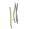
| ||||||||
|---|---|---|---|---|---|---|---|---|---|
| 1 | 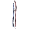
| ||||||||
| 2 | 
| ||||||||
| Unit cell |
|
-Components
| #1: Protein | Mass: 9249.529 Da / Num. of mol.: 2 / Fragment: coiled coil domain Source method: isolated from a genetically manipulated source Source: (gene. exp.) Homo sapiens (human) / Gene: SEPTIN14, SEPT14 / Plasmid: pETSUMO / Production host:  #2: Protein | Mass: 10935.504 Da / Num. of mol.: 2 / Fragment: residues 307-390 Source method: isolated from a genetically manipulated source Source: (gene. exp.) Homo sapiens (human) / Plasmid: pETSUMO / Production host: #3: Chemical | ChemComp-NA /   Mass: 22.990 Da / Num. of mol.: 8 / Source method: obtained synthetically / Formula: Na / Feature type: SUBJECT OF INVESTIGATION Mass: 22.990 Da / Num. of mol.: 8 / Source method: obtained synthetically / Formula: Na / Feature type: SUBJECT OF INVESTIGATION#4: Chemical |   Mass: 106.120 Da / Num. of mol.: 2 / Source method: obtained synthetically / Formula: C4H10O3 / Feature type: SUBJECT OF INVESTIGATION Mass: 106.120 Da / Num. of mol.: 2 / Source method: obtained synthetically / Formula: C4H10O3 / Feature type: SUBJECT OF INVESTIGATION#5: Water | ChemComp-HOH / |  Mass: 18.015 Da / Num. of mol.: 247 / Source method: isolated from a natural source / Formula: H2O Mass: 18.015 Da / Num. of mol.: 247 / Source method: isolated from a natural source / Formula: H2OHas ligand of interest | Y | |
|---|
-Experimental details
-Experiment
| Experiment | Method: X-RAY DIFFRACTION / Number of used crystals: 1 |
|---|
- Sample preparation
| Crystal | Density Matthews: 2.01 Å3/Da / Density % sol: 38.72 % |
|---|---|
| Crystal grow | Temperature: 293 K / Method: vapor diffusion, sitting drop / pH: 5 / Details: 0.1 M Bis-Tris pH 5.0, 24% PEG 3350 |
-Data collection
| Diffraction | Mean temperature: 100 K / Serial crystal experiment: N |
|---|---|
| Diffraction source | Source: SYNCHROTRON / Site: Diamond  / Beamline: I04 / Wavelength: 0.9795 Å / Beamline: I04 / Wavelength: 0.9795 Å |
| Detector | Type: DECTRIS EIGER2 XE 16M / Detector: PIXEL / Date: Feb 25, 2022 |
| Radiation | Protocol: SINGLE WAVELENGTH / Monochromatic (M) / Laue (L): M / Scattering type: x-ray |
| Radiation wavelength | Wavelength: 0.9795 Å / Relative weight: 1 |
| Reflection | Resolution: 1.781→91.294 Å / Num. obs: 22355 / % possible obs: 72.47 % / Redundancy: 6.83 % / CC1/2: 0.999 / Rmerge(I) obs: 0.099 / Rpim(I) all: 0.041 / Rrim(I) all: 0.108 / Net I/σ(I): 10.602 |
| Reflection shell | Resolution: 1.781→1.996 Å / Rmerge(I) obs: 1.168 / Mean I/σ(I) obs: 1.29 / Num. unique obs: 1118 / CC1/2: 0.5692 / Rpim(I) all: 0.535 / Rrim(I) all: 1.1289 / % possible all: 14.18 |
- Processing
| Software |
| |||||||||||||||||||||||||||||||||||||||||||||||||||||||||||||||||||||||||||||||||||||||||||||||||||||||||||||||||||||||||||||
|---|---|---|---|---|---|---|---|---|---|---|---|---|---|---|---|---|---|---|---|---|---|---|---|---|---|---|---|---|---|---|---|---|---|---|---|---|---|---|---|---|---|---|---|---|---|---|---|---|---|---|---|---|---|---|---|---|---|---|---|---|---|---|---|---|---|---|---|---|---|---|---|---|---|---|---|---|---|---|---|---|---|---|---|---|---|---|---|---|---|---|---|---|---|---|---|---|---|---|---|---|---|---|---|---|---|---|---|---|---|---|---|---|---|---|---|---|---|---|---|---|---|---|---|---|---|---|
| Refinement | Method to determine structure: MOLECULAR REPLACEMENT / Resolution: 1.781→91.29 Å / Cor.coef. Fo:Fc: 0.939 / Cor.coef. Fo:Fc free: 0.881 / SU R Cruickshank DPI: 0.228 / Cross valid method: THROUGHOUT / σ(F): 0 / SU R Blow DPI: 0.241 / SU Rfree Blow DPI: 0.208 / SU Rfree Cruickshank DPI: 0.204
| |||||||||||||||||||||||||||||||||||||||||||||||||||||||||||||||||||||||||||||||||||||||||||||||||||||||||||||||||||||||||||||
| Displacement parameters | Biso mean: 32.76 Å2
| |||||||||||||||||||||||||||||||||||||||||||||||||||||||||||||||||||||||||||||||||||||||||||||||||||||||||||||||||||||||||||||
| Refine analyze | Luzzati coordinate error obs: 0.31 Å | |||||||||||||||||||||||||||||||||||||||||||||||||||||||||||||||||||||||||||||||||||||||||||||||||||||||||||||||||||||||||||||
| Refinement step | Cycle: LAST / Resolution: 1.781→91.29 Å
| |||||||||||||||||||||||||||||||||||||||||||||||||||||||||||||||||||||||||||||||||||||||||||||||||||||||||||||||||||||||||||||
| Refine LS restraints |
| |||||||||||||||||||||||||||||||||||||||||||||||||||||||||||||||||||||||||||||||||||||||||||||||||||||||||||||||||||||||||||||
| LS refinement shell | Resolution: 1.781→1.9 Å
| |||||||||||||||||||||||||||||||||||||||||||||||||||||||||||||||||||||||||||||||||||||||||||||||||||||||||||||||||||||||||||||
| Refinement TLS params. | Method: refined / Refine-ID: X-RAY DIFFRACTION
| |||||||||||||||||||||||||||||||||||||||||||||||||||||||||||||||||||||||||||||||||||||||||||||||||||||||||||||||||||||||||||||
| Refinement TLS group |
|
