 Movie
Movie Controller


+ Open data

- Basic information
| Entry | Database: PDB / ID: 8sj3 | ||||||
|---|---|---|---|---|---|---|---|
| Title | Beta-lactamase CTX-M-14 E166Y/N170G | ||||||
 Components Components | Beta-lactamase CTX-M-14 | ||||||
 Keywords Keywords | HYDROLASE / beta-lactamase / enzyme / serine protease / antimicrobial resistance | ||||||
| Function / homology |  Function and homology information Function and homology informationbeta-lactam antibiotic catabolic process / beta-lactamase activity / beta-lactamase / response to antibiotic Similarity search - Function | ||||||
| Biological species |  | ||||||
| Method |  X-RAY DIFFRACTION / SYNCHROTRON / MOLECULAR REPLACEMENT / Resolution: 1.5 Å X-RAY DIFFRACTION / SYNCHROTRON / MOLECULAR REPLACEMENT / Resolution: 1.5 Å | ||||||
 Authors Authors | Judge, A. / Palzkill, T. / Sankaran, B. / Prasad, B.V.V. / Hu, L. | ||||||
| Funding support |  United States, 1items United States, 1items
| ||||||
 Citation Citation | Journal: Proc.Natl.Acad.Sci.USA / Year: 2024 Title: Network of epistatic interactions in an enzyme active site revealed by large-scale deep mutational scanning. Authors: Judge, A. / Sankaran, B. / Hu, L. / Palaniappan, M. / Birgy, A. / Prasad, B.V.V. / Palzkill, T. | ||||||
| History |
|
- Structure visualization
| Structure viewer | Molecule: MolmilJmol/JSmol |
|---|
- Downloads & links
-Download
| PDBx/mmCIF format | 8sj3.cif.gz | 277.5 KB | Display | PDBx/mmCIF format |
|---|---|---|---|---|
| PDB format | pdb8sj3.ent.gz | 186.4 KB | Display | PDB format |
| PDBx/mmJSON format | 8sj3.json.gz | Tree view | PDBx/mmJSON format | |
| Others |  Other downloads Other downloads |
-Validation report
| Summary document | 8sj3_validation.pdf.gz | 431 KB | Display | wwPDB validaton report |
|---|---|---|---|---|
| Full document | 8sj3_full_validation.pdf.gz | 432 KB | Display | |
| Data in XML | 8sj3_validation.xml.gz | 24.3 KB | Display | |
| Data in CIF | 8sj3_validation.cif.gz | 37.1 KB | Display | |
| Arichive directory | https://data.pdbj.org/pub/pdb/validation_reports/sj/8sj3ftp://data.pdbj.org/pub/pdb/validation_reports/sj/8sj3 | HTTPS FTP |
-Related structure data
| Similar structure data |
|---|
-Links
 PDBj
PDBj


- Assembly
| Deposited unit | 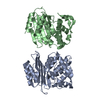
| ||||||||||||
|---|---|---|---|---|---|---|---|---|---|---|---|---|---|
| 1 | 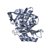
| ||||||||||||
| 2 | 
| ||||||||||||
| Unit cell |
|
-Components
| #1: Protein | Mass: 27977.557 Da / Num. of mol.: 2 Source method: isolated from a genetically manipulated source Source: (gene. exp.) #2: Water | ChemComp-HOH / |  Mass: 18.015 Da / Num. of mol.: 506 / Source method: isolated from a natural source / Formula: H2O Mass: 18.015 Da / Num. of mol.: 506 / Source method: isolated from a natural source / Formula: H2O |
|---|
-Experimental details
-Experiment
| Experiment | Method: X-RAY DIFFRACTION / Number of used crystals: 1 |
|---|
- Sample preparation
| Crystal | Density Matthews: 2.11 Å3/Da / Density % sol: 41.57 % |
|---|---|
| Crystal grow | Temperature: 298 K / Method: vapor diffusion, hanging drop / pH: 5 Details: 0.2 M ammonium chloride, 0.1 M sodium acetate, 20% w/v PEG 6,000 |
-Data collection
| Diffraction | Mean temperature: 100 K / Serial crystal experiment: N |
|---|---|
| Diffraction source | Source: SYNCHROTRON / Site: ALS / Beamline: 8.2.1 / Wavelength: 0.999889 Å |
| Detector | Type: ADSC QUANTUM 315r / Detector: CCD / Date: Apr 28, 2021 |
| Radiation | Monochromator: Double-crystal Si(111) and multilayer / Protocol: SINGLE WAVELENGTH / Monochromatic (M) / Laue (L): M / Scattering type: x-ray |
| Radiation wavelength | Wavelength: 0.999889 Å / Relative weight: 1 |
| Reflection | Resolution: 1.5→32.97 Å / Num. obs: 70681 / % possible obs: 96 % / Redundancy: 4.9 % / Biso Wilson estimate: 13.5 Å2 / CC1/2: 0.82 / Net I/σ(I): 3.26 |
| Reflection shell | Resolution: 1.5→1.554 Å / Mean I/σ(I) obs: 2.18 / Num. unique obs: 7283 / CC1/2: 0.487 |
- Processing
| Software |
| |||||||||||||||||||||||||||||||||||||||||||||||||||||||||||||||||||||||||||||||||||||||||||||||||||||||||||||||||||||||||||||||||||||||||||||||||||||||||||||||||||||||||||||||
|---|---|---|---|---|---|---|---|---|---|---|---|---|---|---|---|---|---|---|---|---|---|---|---|---|---|---|---|---|---|---|---|---|---|---|---|---|---|---|---|---|---|---|---|---|---|---|---|---|---|---|---|---|---|---|---|---|---|---|---|---|---|---|---|---|---|---|---|---|---|---|---|---|---|---|---|---|---|---|---|---|---|---|---|---|---|---|---|---|---|---|---|---|---|---|---|---|---|---|---|---|---|---|---|---|---|---|---|---|---|---|---|---|---|---|---|---|---|---|---|---|---|---|---|---|---|---|---|---|---|---|---|---|---|---|---|---|---|---|---|---|---|---|---|---|---|---|---|---|---|---|---|---|---|---|---|---|---|---|---|---|---|---|---|---|---|---|---|---|---|---|---|---|---|---|---|---|
| Refinement | Method to determine structure: MOLECULAR REPLACEMENT / Resolution: 1.5→32.97 Å / SU ML: 0.1685 / Cross valid method: FREE R-VALUE / σ(F): 1.36 / Phase error: 20.1136 Stereochemistry target values: GeoStd + Monomer Library + CDL v1.2
| |||||||||||||||||||||||||||||||||||||||||||||||||||||||||||||||||||||||||||||||||||||||||||||||||||||||||||||||||||||||||||||||||||||||||||||||||||||||||||||||||||||||||||||||
| Solvent computation | Shrinkage radii: 0.9 Å / VDW probe radii: 1.1 Å / Solvent model: FLAT BULK SOLVENT MODEL | |||||||||||||||||||||||||||||||||||||||||||||||||||||||||||||||||||||||||||||||||||||||||||||||||||||||||||||||||||||||||||||||||||||||||||||||||||||||||||||||||||||||||||||||
| Displacement parameters | Biso mean: 18.07 Å2 | |||||||||||||||||||||||||||||||||||||||||||||||||||||||||||||||||||||||||||||||||||||||||||||||||||||||||||||||||||||||||||||||||||||||||||||||||||||||||||||||||||||||||||||||
| Refinement step | Cycle: LAST / Resolution: 1.5→32.97 Å
| |||||||||||||||||||||||||||||||||||||||||||||||||||||||||||||||||||||||||||||||||||||||||||||||||||||||||||||||||||||||||||||||||||||||||||||||||||||||||||||||||||||||||||||||
| Refine LS restraints |
| |||||||||||||||||||||||||||||||||||||||||||||||||||||||||||||||||||||||||||||||||||||||||||||||||||||||||||||||||||||||||||||||||||||||||||||||||||||||||||||||||||||||||||||||
| LS refinement shell |
|
