 Movie
Movie Controller


+ Open data

- Basic information
| Entry | Database: PDB / ID: 8shy | ||||||
|---|---|---|---|---|---|---|---|
| Title | Structure of binary complex of mouse cGAS QN and bound ATP | ||||||
 Components Components | Cyclic GMP-AMP synthase | ||||||
 Keywords Keywords | TRANSFERASE / IMMUNE SYSTEM | ||||||
| Function / homology |  Function and homology information Function and homology information: / 2',3'-cyclic GMP-AMP synthase activity / cyclic GMP-AMP synthase / regulation of type I interferon production / paracrine signaling / poly-ADP-D-ribose modification-dependent protein binding / regulation of immunoglobulin production / : / regulation of T cell activation / cGAS/STING signaling pathway ...: / 2',3'-cyclic GMP-AMP synthase activity / cyclic GMP-AMP synthase / regulation of type I interferon production / paracrine signaling / poly-ADP-D-ribose modification-dependent protein binding / regulation of immunoglobulin production / : / regulation of T cell activation / cGAS/STING signaling pathway / pattern recognition receptor signaling pathway / negative regulation of DNA repair / negative regulation of cGAS/STING signaling pathway / cellular response to exogenous dsRNA / cytoplasmic pattern recognition receptor signaling pathway / regulation of immune response / negative regulation of double-strand break repair via homologous recombination / nucleosome binding / positive regulation of defense response to virus by host / positive regulation of type I interferon production / phosphatidylinositol-4,5-bisphosphate binding / activation of innate immune response / determination of adult lifespan / molecular condensate scaffold activity / positive regulation of cellular senescence / site of double-strand break / double-stranded DNA binding / defense response to virus / nuclear body / innate immune response / DNA repair / chromatin binding / DNA damage response / GTP binding / protein homodimerization activity / DNA binding / nucleoplasm / ATP binding / metal ion binding / identical protein binding / nucleus / plasma membrane / cytoplasm / cytosol Similarity search - Function | ||||||
| Biological species |  | ||||||
| Method |  X-RAY DIFFRACTION / SYNCHROTRON / MOLECULAR REPLACEMENT / Resolution: 1.77 Å X-RAY DIFFRACTION / SYNCHROTRON / MOLECULAR REPLACEMENT / Resolution: 1.77 Å | ||||||
 Authors Authors | Wu, S. / Sohn, J. | ||||||
| Funding support |  United States, 1items United States, 1items
| ||||||
 Citation Citation | Journal: Nat Commun / Year: 2024 Title: The structural basis for 2'-5'/3'-5'-cGAMP synthesis by cGAS. Authors: Wu, S. / Gabelli, S.B. / Sohn, J. | ||||||
| History |
|
- Structure visualization
| Structure viewer | Molecule: MolmilJmol/JSmol |
|---|
- Downloads & links
-Download
| PDBx/mmCIF format | 8shy.cif.gz | 164.5 KB | Display | PDBx/mmCIF format |
|---|---|---|---|---|
| PDB format | pdb8shy.ent.gz | 127 KB | Display | PDB format |
| PDBx/mmJSON format | 8shy.json.gz | Tree view | PDBx/mmJSON format | |
| Others |  Other downloads Other downloads |
-Validation report
| Arichive directory | https://data.pdbj.org/pub/pdb/validation_reports/sh/8shyftp://data.pdbj.org/pub/pdb/validation_reports/sh/8shy | HTTPS FTP |
|---|
-Related structure data
| Related structure data |  8eaeC  8g10C  8g1jC  8g23C  8gimC 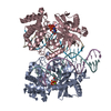 8ginC 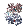 8gioC 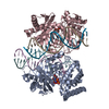 8gipC 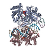 8girC 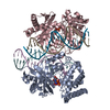 8gisC 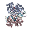 8gitC  8shkC  8shuC  8shzC  8si0C  8sj0C  8sj1C  8sj2C  8sj8C  8sktC  4k8vS S: Starting model for refinement C: citing same article ( |
|---|---|
| Similar structure data |
-Links
 PDBj
PDBj- Assembly
| Deposited unit | 
| ||||||||
|---|---|---|---|---|---|---|---|---|---|
| 1 |
| ||||||||
| Unit cell |
|
-Components
| #1: Protein | Mass: 42638.281 Da / Num. of mol.: 1 / Fragment: catalytic domain (UNP residues 147-507) / Mutation: E211Q, D213N Source method: isolated from a genetically manipulated source Source: (gene. exp.) Details (production host): His*6-MBP-Tev-AgeI-mcGAS CAT, Kanamycin resistance Production host:  |
|---|---|
| #2: Chemical | ChemComp-ZN /   Mass: 65.409 Da / Num. of mol.: 1 / Source method: obtained synthetically / Formula: Zn Mass: 65.409 Da / Num. of mol.: 1 / Source method: obtained synthetically / Formula: Zn |
| #3: Chemical | ChemComp-ATP /   Mass: 507.181 Da / Num. of mol.: 1 / Source method: obtained synthetically / Formula: C10H16N5O13P3 / Feature type: SUBJECT OF INVESTIGATION / Comment: ATP, energy-carrying molecule*YM Mass: 507.181 Da / Num. of mol.: 1 / Source method: obtained synthetically / Formula: C10H16N5O13P3 / Feature type: SUBJECT OF INVESTIGATION / Comment: ATP, energy-carrying molecule*YM |
| #4: Water | ChemComp-HOH /  Mass: 18.015 Da / Num. of mol.: 122 / Source method: isolated from a natural source / Formula: H2O Mass: 18.015 Da / Num. of mol.: 122 / Source method: isolated from a natural source / Formula: H2O |
| Has ligand of interest | Y |
-Experimental details
-Experiment
| Experiment | Method: X-RAY DIFFRACTION / Number of used crystals: 1 |
|---|
- Sample preparation
| Crystal | Density Matthews: 3.28 Å3/Da / Density % sol: 62.46 % |
|---|---|
| Crystal grow | Temperature: 277.15 K / Method: vapor diffusion, hanging drop / pH: 6.5 Details: 0.2 M magnesium acetate tetrahydrate, 0.1 M sodium cacodylate trihydrate, pH 6.5, 20% w/v PEG8000 Temp details: 4-degree Celsius in cold room |
-Data collection
| Diffraction | Mean temperature: 100 K / Ambient temp details: nitrogen gas stream / Serial crystal experiment: N |
|---|---|
| Diffraction source | Source: SYNCHROTRON / Site: NSLS-II / Beamline: 17-ID-1 / Wavelength: 0.9201 Å |
| Detector | Type: DECTRIS EIGER X 9M / Detector: PIXEL / Date: Dec 13, 2021 |
| Radiation | Monochromator: double crystal Si(111) monochromator / Protocol: SINGLE WAVELENGTH / Monochromatic (M) / Laue (L): M / Scattering type: x-ray |
| Radiation wavelength | Wavelength: 0.9201 Å / Relative weight: 1 |
| Reflection | Resolution: 1.77→28.79 Å / Num. obs: 53469 / % possible obs: 99.3 % / Redundancy: 3.5 % / CC1/2: 0.99 / Rmerge(I) obs: 0.083 / Rpim(I) all: 0.052 / Rrim(I) all: 0.098 / Χ2: 1 / Net I/σ(I): 8.1 / Num. measured all: 188329 |
| Reflection shell | Resolution: 1.77→1.8 Å / % possible obs: 97 % / Redundancy: 3.5 % / Rmerge(I) obs: 0.665 / Num. measured all: 10555 / Num. unique obs: 2985 / CC1/2: 0.793 / Rpim(I) all: 0.406 / Rrim(I) all: 0.781 / Χ2: 1.05 / Net I/σ(I) obs: 1.7 |
- Processing
| Software |
| |||||||||||||||||||||||||||||||||||||||||||||||||||||||||||||||||||||||||||||||||||||||||||||||||||||||||
|---|---|---|---|---|---|---|---|---|---|---|---|---|---|---|---|---|---|---|---|---|---|---|---|---|---|---|---|---|---|---|---|---|---|---|---|---|---|---|---|---|---|---|---|---|---|---|---|---|---|---|---|---|---|---|---|---|---|---|---|---|---|---|---|---|---|---|---|---|---|---|---|---|---|---|---|---|---|---|---|---|---|---|---|---|---|---|---|---|---|---|---|---|---|---|---|---|---|---|---|---|---|---|---|---|---|---|
| Refinement | Method to determine structure: MOLECULAR REPLACEMENT Starting model: PDB entry 4K8V Resolution: 1.77→27.94 Å / SU ML: 0.25 / Cross valid method: THROUGHOUT / σ(F): 0.89 / Phase error: 24.88 / Stereochemistry target values: ML
| |||||||||||||||||||||||||||||||||||||||||||||||||||||||||||||||||||||||||||||||||||||||||||||||||||||||||
| Solvent computation | Shrinkage radii: 0.9 Å / VDW probe radii: 1.1 Å / Solvent model: FLAT BULK SOLVENT MODEL | |||||||||||||||||||||||||||||||||||||||||||||||||||||||||||||||||||||||||||||||||||||||||||||||||||||||||
| Refinement step | Cycle: LAST / Resolution: 1.77→27.94 Å
| |||||||||||||||||||||||||||||||||||||||||||||||||||||||||||||||||||||||||||||||||||||||||||||||||||||||||
| Refine LS restraints |
| |||||||||||||||||||||||||||||||||||||||||||||||||||||||||||||||||||||||||||||||||||||||||||||||||||||||||
| LS refinement shell |
| |||||||||||||||||||||||||||||||||||||||||||||||||||||||||||||||||||||||||||||||||||||||||||||||||||||||||
| Refinement TLS params. | Method: refined / Origin x: -3.8406 Å / Origin y: 0.2723 Å / Origin z: 30.3376 Å
| |||||||||||||||||||||||||||||||||||||||||||||||||||||||||||||||||||||||||||||||||||||||||||||||||||||||||
| Refinement TLS group | Selection details: (chain C and resseq 144:507) or (chain A and resseq 2:2) |
