- PDB-8sh1: Structure of human POT1 DNA binding domain bound to a 5'-phosphor... -
+
Open data
ID or keywords:
Loading...
-
Basic information
Entry
Database: PDB / ID: 8sh1
Title
Structure of human POT1 DNA binding domain bound to a 5'-phosphorylated junction of a telomeric double-stranded DNA duplex with a 3'-overhang
Components
DNA (5'-D(*CP*GP*CP*GP*CP*GP*TP*TP*AP*GP*GP*GP*TP*TP*AP*GP*GP*GP*TP*TP*AP*G)-3')
DNA (5'-D(P*CP*TP*AP*AP*CP*GP*CP*GP*CP*G)-3')
Protection of telomeres protein 1
Keywords
DNA BINDING PROTEIN/DNA / shelterin / telomere / DNA junction / 5'-phosphorylated / POT-hole / chromosome end protection / POT1 / DBD / POT1-DNA complex / DNA BINDING PROTEIN / DNA BINDING PROTEIN-DNA complex
Function / homology
Function and homology information
positive regulation of DNA strand elongation / positive regulation of telomeric D-loop disassembly / G-rich single-stranded DNA binding / telomere assembly / 8-hydroxy-2'-deoxyguanosine DNA binding / telomeric D-loop binding / regulation of double-strand break repair via nonhomologous end joining / DEAD/H-box RNA helicase binding / telomerase inhibitor activity / establishment of protein localization to telomere ...positive regulation of DNA strand elongation / positive regulation of telomeric D-loop disassembly / G-rich single-stranded DNA binding / telomere assembly / 8-hydroxy-2'-deoxyguanosine DNA binding / telomeric D-loop binding / regulation of double-strand break repair via nonhomologous end joining / DEAD/H-box RNA helicase binding / telomerase inhibitor activity / establishment of protein localization to telomere / telomeric D-loop disassembly / shelterin complex / Telomere C-strand synthesis initiation / regulation of telomere maintenance via telomerase / Telomere C-strand (Lagging Strand) Synthesis / nuclear telomere cap complex / single-stranded telomeric DNA binding / G-rich strand telomeric DNA binding / telomere capping / Processive synthesis on the C-strand of the telomere / Polymerase switching on the C-strand of the telomere / Removal of the Flap Intermediate from the C-strand / telomeric DNA binding / negative regulation of telomere maintenance via telomerase / positive regulation of telomere maintenance / Telomere Extension By Telomerase / telomere maintenance via telomerase / Packaging Of Telomere Ends / positive regulation of telomere maintenance via telomerase / Recognition and association of DNA glycosylase with site containing an affected purine / Cleavage of the damaged purine / Recognition and association of DNA glycosylase with site containing an affected pyrimidine / Cleavage of the damaged pyrimidine / Inhibition of DNA recombination at telomere / Meiotic synapsis / DNA Damage/Telomere Stress Induced Senescence / chromosome, telomeric region / nucleoplasm Similarity search - Function
: / Protection of telomeres protein 1 (POT1), C-terminal insertion domain / Protection of telomeres protein 1, ssDNA-binding domain / ssDNA-binding domain of telomere protection protein / Protection of telomeres protein 1 / Telomeric single stranded DNA binding POT1/Cdc13 / Telomeric single stranded DNA binding POT1/CDC13 / Telomeric single stranded DNA binding POT1/CDC13 / Nucleic acid-binding proteins / OB fold (Dihydrolipoamide Acetyltransferase, E2P) ...: / Protection of telomeres protein 1 (POT1), C-terminal insertion domain / Protection of telomeres protein 1, ssDNA-binding domain / ssDNA-binding domain of telomere protection protein / Protection of telomeres protein 1 / Telomeric single stranded DNA binding POT1/Cdc13 / Telomeric single stranded DNA binding POT1/CDC13 / Telomeric single stranded DNA binding POT1/CDC13 / Nucleic acid-binding proteins / OB fold (Dihydrolipoamide Acetyltransferase, E2P) / Nucleic acid-binding, OB-fold / Beta Barrel / Mainly Beta Similarity search - Domain/homology
#236 - Aug 2019 Cyclin and Cyclin-dependent Kinase similarity (1)
-
Assembly
Deposited unit
A: Protection of telomeres protein 1 B: DNA (5'-D(*CP*GP*CP*GP*CP*GP*TP*TP*AP*GP*GP*GP*TP*TP*AP*GP*GP*GP*TP*TP*AP*G)-3') C: DNA (5'-D(P*CP*TP*AP*AP*CP*GP*CP*GP*CP*G)-3')
Protectionoftelomeresprotein1 / hPot1 / POT1-like telomere end-binding protein
Mass: 33811.828 Da / Num. of mol.: 1 / Fragment: DNA binding domain Source method: isolated from a genetically manipulated source Source: (gene. exp.) Homo sapiens (human) / Gene: POT1 / Plasmid: pFBHTb-Smt3star-hPOT1-1-299 Details (production host): human POT1 aa 1-299 cloned downstream of a sumo-star tag in the pFastBac vector with an N-terminal His tag Cell line (production host): High Five / Production host: Trichoplusia ni (cabbage looper) / References: UniProt: Q9NUX5
#2: DNA chain
DNA (5'-D(*CP*GP*CP*GP*CP*GP*TP*TP*AP*GP*GP*GP*TP*TP*AP*GP*GP*GP*TP*TP*AP*G)-3')
Mass: 6879.424 Da / Num. of mol.: 1 / Source method: obtained synthetically / Source: (synth.) Homo sapiens (human)
#3: DNA chain
DNA (5'-D(P*CP*TP*AP*AP*CP*GP*CP*GP*CP*G)-3')
Mass: 3029.993 Da / Num. of mol.: 1 / Source method: obtained synthetically / Source: (synth.) Homo sapiens (human)
Mass: 18.015 Da / Num. of mol.: 45 / Source method: isolated from a natural source / Formula: H2O
-
Experimental details
-
Experiment
Experiment
Method: X-RAY DIFFRACTION / Number of used crystals: 1
-
Sample preparation
Crystal
Density Matthews: 4.96 Å3/Da / Density % sol: 75.21 %
Crystal grow
Temperature: 289 K / Method: vapor diffusion, sitting drop / pH: 8 Details: hDBD complex with 5prime-P-ds-ss1-12 was crystallized at 16C by the sitting drop method in a drop containing 0.5 microliter 14 mg/ml protein-DNA complex (in 25 mM Tris (pH 8), 100 mM NaCl, ...Details: hDBD complex with 5prime-P-ds-ss1-12 was crystallized at 16C by the sitting drop method in a drop containing 0.5 microliter 14 mg/ml protein-DNA complex (in 25 mM Tris (pH 8), 100 mM NaCl, and 2 mM DTT) and 0.5 microliter well solution (0.02 M MgCl2, 0.9 M Hepes (pH 7.5), and 20% polyacrylic acid N100 sodium salt, derived from condition G2 of the JCSG+ screen (NeXtal). Crystals were harvested in 25 mM Tris (pH 8), 100 mM NaCl, 0.02 M MgCl2, 0.1 M Hepes (pH 7.5), and 22% polyacrylic acid N100 sodium salt and cryoprotected in harvesting solution supplemented with 35% ethylene glycol
-
Data collection
Diffraction
Mean temperature: 100 K / Serial crystal experiment: N
In the structure databanks used in Yorodumi, some data are registered as the other names, "COVID-19 virus" and "2019-nCoV". Here are the details of the virus and the list of structure data.
Jan 31, 2019. EMDB accession codes are about to change! (news from PDBe EMDB page)
EMDB accession codes are about to change! (news from PDBe EMDB page)
The allocation of 4 digits for EMDB accession codes will soon come to an end. Whilst these codes will remain in use, new EMDB accession codes will include an additional digit and will expand incrementally as the available range of codes is exhausted. The current 4-digit format prefixed with “EMD-” (i.e. EMD-XXXX) will advance to a 5-digit format (i.e. EMD-XXXXX), and so on. It is currently estimated that the 4-digit codes will be depleted around Spring 2019, at which point the 5-digit format will come into force.
The EM Navigator/Yorodumi systems omit the EMD- prefix.
Related info.:Q: What is EMD? / ID/Accession-code notation in Yorodumi/EM Navigator
Yorodumi is a browser for structure data from EMDB, PDB, SASBDB, etc.
This page is also the successor to EM Navigator detail page, and also detail information page/front-end page for Omokage search.
The word "yorodu" (or yorozu) is an old Japanese word meaning "ten thousand". "mi" (miru) is to see.
Related info.:EMDB / PDB / SASBDB / Comparison of 3 databanks / Yorodumi Search / Aug 31, 2016. New EM Navigator & Yorodumi / Yorodumi Papers / Jmol/JSmol / Function and homology information / Changes in new EM Navigator and Yorodumi
 Movie
Movie Controller

 Yorodumi
 Open data
 Basic information
 Components
 Keywords
 Function and homology information
 Homo sapiens (human)
 X-RAY DIFFRACTION /
  Authors
 United States, 4items
  Citation
 Structure visualization
 Downloads & links
 Other downloads
 PDBj
PDBj


 Assembly
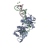
 Trichoplusia ni (cabbage looper) / References: UniProt: Q9NUX5
 Mass: 18.015 Da / Num. of mol.: 45 / Source method: isolated from a natural source / Formula: H2O
 Sample preparation
 Processing