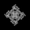
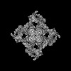
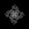
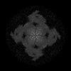
 Movie
Movie Controller


+ Open data

- Basic information
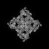
| Entry | Database: PDB / ID: 8seu | |||||||||
|---|---|---|---|---|---|---|---|---|---|---|
| Title | Cryo-EM Structure of RyR1 (Local Refinement of TMD) | |||||||||
 Components Components | Ryanodine receptor 1 | |||||||||
 Keywords Keywords | TRANSPORT PROTEIN / Calcium ion channel / skeletal muscle / nucleotide / homotetramer | |||||||||
| Function / homology |  Function and homology information Function and homology informationATP-gated ion channel activity / ryanodine-sensitive calcium-release channel activity / terminal cisterna / ryanodine receptor complex / release of sequestered calcium ion into cytosol by sarcoplasmic reticulum / ossification involved in bone maturation / cellular response to caffeine / skin development / organelle membrane / smooth endoplasmic reticulum ...ATP-gated ion channel activity / ryanodine-sensitive calcium-release channel activity / terminal cisterna / ryanodine receptor complex / release of sequestered calcium ion into cytosol by sarcoplasmic reticulum / ossification involved in bone maturation / cellular response to caffeine / skin development / organelle membrane / smooth endoplasmic reticulum / intracellularly gated calcium channel activity / outflow tract morphogenesis / toxic substance binding / voltage-gated calcium channel activity / striated muscle contraction / skeletal muscle fiber development / release of sequestered calcium ion into cytosol / sarcoplasmic reticulum membrane / cellular response to calcium ion / muscle contraction / sarcoplasmic reticulum / sarcolemma / Z disc / intracellular calcium ion homeostasis / calcium ion transmembrane transport / calcium channel activity / disordered domain specific binding / protein homotetramerization / transmembrane transporter binding / calmodulin binding / calcium ion binding / ATP binding / membrane / identical protein binding Similarity search - Function | |||||||||
| Biological species |  | |||||||||
| Method | ELECTRON MICROSCOPY / single particle reconstruction / cryo EM / Resolution: 3 Å | |||||||||
 Authors Authors | Cholak, S. / Saville, J.W. / Zhu, X. / Berezuk, A.M. / Tuttle, K.S. / Haji-Ghassemi, O. / Van Petegem, F. / Subramaniam, S. | |||||||||
| Funding support |  Canada, 2items Canada, 2items
| |||||||||
 Citation Citation | Journal: Structure / Year: 2023 Title: Allosteric modulation of ryanodine receptor RyR1 by nucleotide derivatives. Authors: Spencer Cholak / James W Saville / Xing Zhu / Alison M Berezuk / Katharine S Tuttle / Omid Haji-Ghassemi / Francisco J Alvarado / Filip Van Petegem / Sriram Subramaniam /  Abstract: The coordinated release of Ca from the sarcoplasmic reticulum (SR) is critical for excitation-contraction coupling. This release is facilitated by ryanodine receptors (RyRs) that are embedded in the ...The coordinated release of Ca from the sarcoplasmic reticulum (SR) is critical for excitation-contraction coupling. This release is facilitated by ryanodine receptors (RyRs) that are embedded in the SR membrane. In skeletal muscle, activity of RyR1 is regulated by metabolites such as ATP, which upon binding increase channel open probability (P). To obtain structural insights into the mechanism of RyR1 priming by ATP, we determined several cryo-EM structures of RyR1 bound individually to ATP-γ-S, ADP, AMP, adenosine, adenine, and cAMP. We demonstrate that adenine and adenosine bind RyR1, but AMP is the smallest ATP derivative capable of inducing long-range (>170 Å) structural rearrangements associated with channel activation, establishing a structural basis for key binding site interactions that are the threshold for triggering quaternary structural changes. Our finding that cAMP also induces these structural changes and results in increased channel opening suggests its potential role as an endogenous modulator of RyR1 conductance. | |||||||||
| History |
|
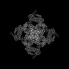
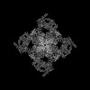
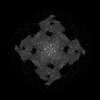
- Structure visualization
| Structure viewer | Molecule: MolmilJmol/JSmol |
|---|
- Downloads & links
-Download
| PDBx/mmCIF format | 8seu.cif.gz | 729.5 KB | Display | PDBx/mmCIF format |
|---|---|---|---|---|
| PDB format | pdb8seu.ent.gz | 438 KB | Display | PDB format |
| PDBx/mmJSON format | 8seu.json.gz | Tree view | PDBx/mmJSON format | |
| Others |  Other downloads Other downloads |
-Validation report
| Arichive directory | https://data.pdbj.org/pub/pdb/validation_reports/se/8seuftp://data.pdbj.org/pub/pdb/validation_reports/se/8seu | HTTPS FTP |
|---|
-Related structure data
| Related structure data | 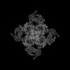 40429MC  8senC 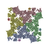 8seoC  8sepC  8seqC  8serC  8sesC  8setC 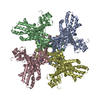 8sevC  8sewC  8sexC  8seyC  8sezC  8sf0C M: map data used to model this data C: citing same article ( |
|---|---|
| Similar structure data |
-Links
 PDBj
PDBj
- Assembly
| Deposited unit | 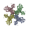
|
|---|---|
| 1 |
|
-Components
| #1: Protein | Mass: 565908.625 Da / Num. of mol.: 4 / Source method: isolated from a natural source / Source: (natural) #2: Chemical | ChemComp-ZN /   Mass: 65.409 Da / Num. of mol.: 4 / Source method: obtained synthetically / Formula: Zn Mass: 65.409 Da / Num. of mol.: 4 / Source method: obtained synthetically / Formula: ZnHas ligand of interest | N | Has protein modification | Y | |
|---|
-Experimental details
-Experiment
| Experiment | Method: ELECTRON MICROSCOPY |
|---|---|
| EM experiment | Aggregation state: PARTICLE / 3D reconstruction method: single particle reconstruction |
- Sample preparation
| Component | Name: RyR1 in complex with FKBP12.6 / Type: COMPLEX / Entity ID: #1 / Source: MULTIPLE SOURCES |
|---|---|
| Source (natural) | Organism: |
| Buffer solution | pH: 7.5 |
| Specimen | Embedding applied: NO / Shadowing applied: NO / Staining applied: NO / Vitrification applied: YES |
| Specimen support | Grid material: COPPER / Grid mesh size: 300 divisions/in. / Grid type: Quantifoil R1.2/1.3 |
| Vitrification | Instrument: FEI VITROBOT MARK IV / Cryogen name: ETHANE / Humidity: 100 % / Chamber temperature: 283.15 K |
- Electron microscopy imaging
| Experimental equipment |  Model: Titan Krios / Image courtesy: FEI Company |
|---|---|
| Microscopy | Model: TFS KRIOS |
| Electron gun | Electron source:  FIELD EMISSION GUN / Accelerating voltage: 300 kV / Illumination mode: FLOOD BEAM FIELD EMISSION GUN / Accelerating voltage: 300 kV / Illumination mode: FLOOD BEAM |
| Electron lens | Mode: BRIGHT FIELD / Nominal magnification: 96000 X / Nominal defocus max: 2000 nm / Nominal defocus min: 500 nm / Cs: 2.7 mm / C2 aperture diameter: 50 µm / Alignment procedure: COMA FREE |
| Specimen holder | Cryogen: NITROGEN / Specimen holder model: FEI TITAN KRIOS AUTOGRID HOLDER |
| Image recording | Electron dose: 40 e/Å2 / Film or detector model: FEI FALCON IV (4k x 4k) |
- Processing
| EM software | Name: EPU / Version: 3.3 / Category: image acquisition |
|---|---|
| CTF correction | Type: PHASE FLIPPING AND AMPLITUDE CORRECTION |
| 3D reconstruction | Resolution: 3 Å / Resolution method: FSC 0.143 CUT-OFF / Num. of particles: 45876 / Symmetry type: POINT |
