+ Open data
- Basic information
| Entry | Database: PDB / ID: 8sd3 | ||||||
|---|---|---|---|---|---|---|---|
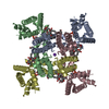
| Title | CryoEM structure of rat Kv2.1(1-598) wild type in nanodiscs | ||||||
 Components Components | Potassium voltage-gated channel subfamily B member 1 | ||||||
 Keywords Keywords | MEMBRANE PROTEIN / voltage-dependent potassium channel | ||||||
| Function / homology |  Function and homology information Function and homology informationregulation of action potential / regulation of motor neuron apoptotic process / Voltage gated Potassium channels / clustering of voltage-gated potassium channels / positive regulation of long-term synaptic depression / positive regulation of norepinephrine secretion / positive regulation of catecholamine secretion / cholinergic synapse / potassium ion export across plasma membrane / positive regulation of calcium ion-dependent exocytosis ...regulation of action potential / regulation of motor neuron apoptotic process / Voltage gated Potassium channels / clustering of voltage-gated potassium channels / positive regulation of long-term synaptic depression / positive regulation of norepinephrine secretion / positive regulation of catecholamine secretion / cholinergic synapse / potassium ion export across plasma membrane / positive regulation of calcium ion-dependent exocytosis / proximal dendrite / delayed rectifier potassium channel activity / outward rectifier potassium channel activity / Glucagon-like Peptide-1 (GLP1) regulates insulin secretion / response to L-glutamate / vesicle docking involved in exocytosis / postsynaptic specialization membrane / glutamate receptor signaling pathway / neuronal cell body membrane / action potential / voltage-gated potassium channel activity / lateral plasma membrane / positive regulation of protein targeting to membrane / potassium channel regulator activity / response to axon injury / cellular response to nutrient levels / voltage-gated potassium channel complex / potassium ion transmembrane transport / dendrite membrane / cellular response to calcium ion / SNARE binding / protein localization to plasma membrane / cellular response to glucose stimulus / negative regulation of insulin secretion / protein homooligomerization / sarcolemma / potassium ion transport / glucose homeostasis / perikaryon / postsynaptic membrane / transmembrane transporter binding / apical plasma membrane / protein heterodimerization activity / axon / neuronal cell body / dendrite / perinuclear region of cytoplasm / cell surface / plasma membrane Similarity search - Function | ||||||
| Biological species |  | ||||||
| Method | ELECTRON MICROSCOPY / single particle reconstruction / cryo EM / Resolution: 2.95 Å | ||||||
 Authors Authors | Tan, X. / Swartz, K.J. | ||||||
| Funding support |  United States, 1items United States, 1items
| ||||||
 Citation Citation |  Journal: Nature / Year: 2023 Journal: Nature / Year: 2023Title: Inactivation of the Kv2.1 channel through electromechanical coupling. Authors: Ana I Fernández-Mariño / Xiao-Feng Tan / Chanhyung Bae / Kate Huffer / Jiansen Jiang / Kenton J Swartz /  Abstract: The Kv2.1 voltage-activated potassium (Kv) channel is a prominent delayed-rectifier Kv channel in the mammalian central nervous system, where its mechanisms of activation and inactivation are ...The Kv2.1 voltage-activated potassium (Kv) channel is a prominent delayed-rectifier Kv channel in the mammalian central nervous system, where its mechanisms of activation and inactivation are critical for regulating intrinsic neuronal excitability. Here we present structures of the Kv2.1 channel in a lipid environment using cryo-electron microscopy to provide a framework for exploring its functional mechanisms and how mutations causing epileptic encephalopathies alter channel activity. By studying a series of disease-causing mutations, we identified one that illuminates a hydrophobic coupling nexus near the internal end of the pore that is critical for inactivation. Both functional and structural studies reveal that inactivation in Kv2.1 results from dynamic alterations in electromechanical coupling to reposition pore-lining S6 helices and close the internal pore. Consideration of these findings along with available structures for other Kv channels, as well as voltage-activated sodium and calcium channels, suggests that related mechanisms of inactivation are conserved in voltage-activated cation channels and likely to be engaged by widely used therapeutics to achieve state-dependent regulation of channel activity. | ||||||
| History |
|
- Structure visualization
| Structure viewer | Molecule:  Molmil Molmil Jmol/JSmol Jmol/JSmol |
|---|
- Downloads & links
- Download
| PDBx/mmCIF format |  8sd3.cif.gz 8sd3.cif.gz | 342.1 KB | Display |  PDBx/mmCIF format PDBx/mmCIF format |
|---|---|---|---|---|
| PDB format |  pdb8sd3.ent.gz pdb8sd3.ent.gz | 262.8 KB | Display |  PDB format PDB format |
| PDBx/mmJSON format |  8sd3.json.gz 8sd3.json.gz | Tree view |  PDBx/mmJSON format PDBx/mmJSON format | |
| Others |  Other downloads Other downloads |
-Validation report
| Summary document |  8sd3_validation.pdf.gz 8sd3_validation.pdf.gz | 1.7 MB | Display |  wwPDB validaton report wwPDB validaton report |
|---|---|---|---|---|
| Full document |  8sd3_full_validation.pdf.gz 8sd3_full_validation.pdf.gz | 1.7 MB | Display | |
| Data in XML |  8sd3_validation.xml.gz 8sd3_validation.xml.gz | 42.1 KB | Display | |
| Data in CIF |  8sd3_validation.cif.gz 8sd3_validation.cif.gz | 53 KB | Display | |
| Arichive directory |  https://data.pdbj.org/pub/pdb/validation_reports/sd/8sd3 https://data.pdbj.org/pub/pdb/validation_reports/sd/8sd3 ftp://data.pdbj.org/pub/pdb/validation_reports/sd/8sd3 ftp://data.pdbj.org/pub/pdb/validation_reports/sd/8sd3 | HTTPS FTP |
-Related structure data
| Related structure data |  40349MC  8sdaC M: map data used to model this data C: citing same article ( |
|---|---|
| Similar structure data | Similarity search - Function & homology  F&H Search F&H Search |
- Links
Links
- Assembly
| Deposited unit | 
|
|---|---|
| 1 |
|
- Components
| #1: Protein | Mass: 68592.102 Da / Num. of mol.: 4 Source method: isolated from a genetically manipulated source Source: (gene. exp.)   Homo sapiens (human) / Strain (production host): TSA201 / References: UniProt: P15387 Homo sapiens (human) / Strain (production host): TSA201 / References: UniProt: P15387#2: Chemical | ChemComp-POV / ( #3: Chemical | ChemComp-K / Has ligand of interest | N | |
|---|
-Experimental details
-Experiment
| Experiment | Method: ELECTRON MICROSCOPY |
|---|---|
| EM experiment | Aggregation state: PARTICLE / 3D reconstruction method: single particle reconstruction |
- Sample preparation
| Component | Name: Voltage-dependent potassium channel Kv2.1 / Type: COMPLEX / Entity ID: #1 / Source: RECOMBINANT |
|---|---|
| Source (natural) | Organism:  |
| Source (recombinant) | Organism:  Homo sapiens (human) / Cell: tsa201 Homo sapiens (human) / Cell: tsa201 |
| Buffer solution | pH: 7.5 |
| Specimen | Embedding applied: NO / Shadowing applied: NO / Staining applied: NO / Vitrification applied: YES |
| Specimen support | Grid material: COPPER / Grid mesh size: 300 divisions/in. / Grid type: Quantifoil R1.2/1.3 |
| Vitrification | Instrument: FEI VITROBOT MARK IV / Cryogen name: ETHANE / Humidity: 100 % |
- Electron microscopy imaging
| Experimental equipment |  Model: Titan Krios / Image courtesy: FEI Company |
|---|---|
| Microscopy | Model: FEI TITAN KRIOS |
| Electron gun | Electron source: TUNGSTEN HAIRPIN / Accelerating voltage: 300 kV / Illumination mode: FLOOD BEAM |
| Electron lens | Mode: BRIGHT FIELD / Nominal defocus max: 2000 nm / Nominal defocus min: 500 nm |
| Image recording | Electron dose: 71 e/Å2 / Film or detector model: GATAN K2 SUMMIT (4k x 4k) |
- Processing
| Software |
| ||||||||||||||||||||||||
|---|---|---|---|---|---|---|---|---|---|---|---|---|---|---|---|---|---|---|---|---|---|---|---|---|---|
| CTF correction | Type: NONE | ||||||||||||||||||||||||
| 3D reconstruction | Resolution: 2.95 Å / Resolution method: FSC 0.143 CUT-OFF / Num. of particles: 73548 / Symmetry type: POINT | ||||||||||||||||||||||||
| Atomic model building | Protocol: RIGID BODY FIT | ||||||||||||||||||||||||
| Refinement | Cross valid method: NONE Stereochemistry target values: GeoStd + Monomer Library + CDL v1.2 | ||||||||||||||||||||||||
| Displacement parameters | Biso mean: 64.33 Å2 | ||||||||||||||||||||||||
| Refine LS restraints |
|
 Movie
Movie Controller


 PDBj
PDBj


 gel filtration


