 Movie
Movie Controller

[English] 日本語
 Yorodumi
- PDB-8sbo: Crystal Structure of Dephospho-CoA kinase from Klebsiella aerogen... -
+ Open data

- Basic information
| Entry | Database: PDB / ID: 8sbo | |||||||||
|---|---|---|---|---|---|---|---|---|---|---|
| Title | Crystal Structure of Dephospho-CoA kinase from Klebsiella aerogenes (P21 Form 2) | |||||||||
 Components Components | Dephospho-CoA kinase | |||||||||
 Keywords Keywords | TRANSFERASE / SSGCID / STRUCTURAL GENOMICS / SEATTLE STRUCTURAL GENOMICS CENTER FOR INFECTIOUS DISEASE | |||||||||
| Function / homology |  Function and homology information Function and homology informationdephospho-CoA kinase / dephospho-CoA kinase activity / coenzyme A biosynthetic process / ATP binding / cytoplasm Similarity search - Function | |||||||||
| Biological species |  Klebsiella aerogenes KCTC 2190 (bacteria) Klebsiella aerogenes KCTC 2190 (bacteria) | |||||||||
| Method |  X-RAY DIFFRACTION / SYNCHROTRON / MOLECULAR REPLACEMENT / Resolution: 1.4 Å X-RAY DIFFRACTION / SYNCHROTRON / MOLECULAR REPLACEMENT / Resolution: 1.4 Å | |||||||||
 Authors Authors | Seattle Structural Genomics Center for Infectious Disease (SSGCID) | |||||||||
| Funding support |  United States, 2items United States, 2items
| |||||||||
 Citation Citation | Journal: To be published Title: Crystal Structure of Dephospho-CoA kinase from Klebsiella aerogenes (P21 Form 2) Authors: Liu, L. / Lovell, S. / Battaile, K.P. / Seibold, S. | |||||||||
| History |
|
- Structure visualization
| Structure viewer | Molecule: MolmilJmol/JSmol |
|---|
- Downloads & links
-Download
| PDBx/mmCIF format | 8sbo.cif.gz | 180 KB | Display | PDBx/mmCIF format |
|---|---|---|---|---|
| PDB format | pdb8sbo.ent.gz | 141.5 KB | Display | PDB format |
| PDBx/mmJSON format | 8sbo.json.gz | Tree view | PDBx/mmJSON format | |
| Others |  Other downloads Other downloads |
-Validation report
| Arichive directory | https://data.pdbj.org/pub/pdb/validation_reports/sb/8sboftp://data.pdbj.org/pub/pdb/validation_reports/sb/8sbo | HTTPS FTP |
|---|
-Related structure data
| Similar structure data |
|---|
-Links
 PDBj
PDBj- Assembly
| Deposited unit | 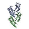
| ||||||||
|---|---|---|---|---|---|---|---|---|---|
| 1 | 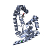
| ||||||||
| 2 | 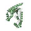
| ||||||||
| Unit cell |
|
-Components
| #1: Protein | Mass: 23850.084 Da / Num. of mol.: 2 / Mutation: G2T, V127I Source method: isolated from a genetically manipulated source Source: (gene. exp.) Klebsiella aerogenes KCTC 2190 (bacteria)Gene: coaE, EAE_11320 / Plasmid: KlaeA.00139.a.B1 / Production host: #2: Chemical |   Mass: 35.453 Da / Num. of mol.: 2 / Source method: obtained synthetically / Formula: Cl Mass: 35.453 Da / Num. of mol.: 2 / Source method: obtained synthetically / Formula: Cl#3: Chemical |   Mass: 118.174 Da / Num. of mol.: 2 / Source method: isolated from a natural source / Formula: C6H14O2 / Comment: precipitant*YM Mass: 118.174 Da / Num. of mol.: 2 / Source method: isolated from a natural source / Formula: C6H14O2 / Comment: precipitant*YM#4: Water | ChemComp-HOH / |  Mass: 18.015 Da / Num. of mol.: 350 / Source method: isolated from a natural source / Formula: H2O Mass: 18.015 Da / Num. of mol.: 350 / Source method: isolated from a natural source / Formula: H2OHas ligand of interest | N | |
|---|
-Experimental details
-Experiment
| Experiment | Method: X-RAY DIFFRACTION / Number of used crystals: 1 |
|---|
- Sample preparation
| Crystal | Density Matthews: 2.08 Å3/Da / Density % sol: 41 % |
|---|---|
| Crystal grow | Temperature: 291 K / Method: vapor diffusion, sitting drop / pH: 6.5 Details: Morpheus H4: 12.5%(v/v) MPD, 12.5%(v/v) PEG 1000, 12.5%(w/v) PEG 3350, 100 mM Imidazole/MES, pH 6.5, 20 mM DL-Glutamic acid, 20 mM DL-Alanine; 20 mM Glycine, 20 mM DL-Lysine ...Details: Morpheus H4: 12.5%(v/v) MPD, 12.5%(v/v) PEG 1000, 12.5%(w/v) PEG 3350, 100 mM Imidazole/MES, pH 6.5, 20 mM DL-Glutamic acid, 20 mM DL-Alanine; 20 mM Glycine, 20 mM DL-Lysine monohydrochloride and 20 mM DL-Serine. KlaeA.00139.a.B1.PW39166 at 24.8 mg/mL. Plate: 13153, well H4 drop 1, Puck: PSL-1416, Cryo: Direct |
-Data collection
| Diffraction | Mean temperature: 100 K / Serial crystal experiment: N |
|---|---|
| Diffraction source | Source: SYNCHROTRON / Site: NSLS-II / Beamline: 19-ID / Wavelength: 0.9795 Å |
| Detector | Type: DECTRIS EIGER2 XE 9M / Detector: PIXEL / Date: Feb 14, 2022 |
| Radiation | Monochromator: Double Crystal Si 111 / Protocol: SINGLE WAVELENGTH / Monochromatic (M) / Laue (L): M / Scattering type: x-ray |
| Radiation wavelength | Wavelength: 0.9795 Å / Relative weight: 1 |
| Reflection | Resolution: 1.4→63.81 Å / Num. obs: 77530 / % possible obs: 100 % / Redundancy: 6.8 % / CC1/2: 1 / Rmerge(I) obs: 0.043 / Rpim(I) all: 0.017 / Rrim(I) all: 0.047 / Χ2: 0.97 / Net I/σ(I): 16.2 / Num. measured all: 529323 |
| Reflection shell | Resolution: 1.4→1.44 Å / % possible obs: 100 % / Redundancy: 6.8 % / Rmerge(I) obs: 1.121 / Num. measured all: 38647 / Num. unique obs: 5715 / CC1/2: 0.774 / Rpim(I) all: 0.461 / Rrim(I) all: 1.214 / Χ2: 0.98 / Net I/σ(I) obs: 1.5 |
- Processing
| Software |
| ||||||||||||||||||||||||||||||||||||||||||||||||||||||||||||||||||||||||||||||||||||||||||||||||||||||||||||||||||||||||||||||||||||||||||||||||||||||||||||||||||||||||||||||||||||||||||||||||||||
|---|---|---|---|---|---|---|---|---|---|---|---|---|---|---|---|---|---|---|---|---|---|---|---|---|---|---|---|---|---|---|---|---|---|---|---|---|---|---|---|---|---|---|---|---|---|---|---|---|---|---|---|---|---|---|---|---|---|---|---|---|---|---|---|---|---|---|---|---|---|---|---|---|---|---|---|---|---|---|---|---|---|---|---|---|---|---|---|---|---|---|---|---|---|---|---|---|---|---|---|---|---|---|---|---|---|---|---|---|---|---|---|---|---|---|---|---|---|---|---|---|---|---|---|---|---|---|---|---|---|---|---|---|---|---|---|---|---|---|---|---|---|---|---|---|---|---|---|---|---|---|---|---|---|---|---|---|---|---|---|---|---|---|---|---|---|---|---|---|---|---|---|---|---|---|---|---|---|---|---|---|---|---|---|---|---|---|---|---|---|---|---|---|---|---|---|---|---|
| Refinement | Method to determine structure: MOLECULAR REPLACEMENT / Resolution: 1.4→21.17 Å / SU ML: 0.16 / Cross valid method: FREE R-VALUE / σ(F): 1.97 / Phase error: 22.59 / Stereochemistry target values: ML
| ||||||||||||||||||||||||||||||||||||||||||||||||||||||||||||||||||||||||||||||||||||||||||||||||||||||||||||||||||||||||||||||||||||||||||||||||||||||||||||||||||||||||||||||||||||||||||||||||||||
| Solvent computation | Shrinkage radii: 0.9 Å / VDW probe radii: 1.1 Å / Solvent model: FLAT BULK SOLVENT MODEL | ||||||||||||||||||||||||||||||||||||||||||||||||||||||||||||||||||||||||||||||||||||||||||||||||||||||||||||||||||||||||||||||||||||||||||||||||||||||||||||||||||||||||||||||||||||||||||||||||||||
| Refinement step | Cycle: LAST / Resolution: 1.4→21.17 Å
| ||||||||||||||||||||||||||||||||||||||||||||||||||||||||||||||||||||||||||||||||||||||||||||||||||||||||||||||||||||||||||||||||||||||||||||||||||||||||||||||||||||||||||||||||||||||||||||||||||||
| Refine LS restraints |
| ||||||||||||||||||||||||||||||||||||||||||||||||||||||||||||||||||||||||||||||||||||||||||||||||||||||||||||||||||||||||||||||||||||||||||||||||||||||||||||||||||||||||||||||||||||||||||||||||||||
| LS refinement shell |
|
