 Movie
Movie Controller

[English] 日本語
 Yorodumi
- PDB-8sa9: Crystal Structure of Cystathionine beta lyase from Klebsiella aer... -
+ Open data

- Basic information
| Entry | Database: PDB / ID: 8sa9 | |||||||||
|---|---|---|---|---|---|---|---|---|---|---|
| Title | Crystal Structure of Cystathionine beta lyase from Klebsiella aerogenes, PLP-Oxamate Adduct (C2 form) | |||||||||
 Components Components | Cystathionine beta-lyase | |||||||||
 Keywords Keywords | LYASE / SSGCID / STRUCTURAL GENOMICS / SEATTLE STRUCTURAL GENOMICS CENTER FOR INFECTIOUS DISEASE | |||||||||
| Function / homology |  Function and homology information Function and homology information: / cysteine-S-conjugate beta-lyase activity / cysteine-S-conjugate beta-lyase / transsulfuration / : / pyridoxal phosphate binding / metal ion binding / cytoplasm Similarity search - Function | |||||||||
| Biological species |  Klebsiella aerogenes KCTC 2190 (bacteria) Klebsiella aerogenes KCTC 2190 (bacteria) | |||||||||
| Method |  X-RAY DIFFRACTION / SYNCHROTRON / MOLECULAR REPLACEMENT / Resolution: 1.1 Å X-RAY DIFFRACTION / SYNCHROTRON / MOLECULAR REPLACEMENT / Resolution: 1.1 Å | |||||||||
 Authors Authors | Seattle Structural Genomics Center for Infectious Disease (SSGCID) | |||||||||
| Funding support |  United States, 2items United States, 2items
| |||||||||
 Citation Citation | Journal: To be published Title: Crystal Structure of Cystathionine beta lyase from Klebsiella aerogenes, PLP-Oxamate Adduct (C2 form) Authors: Liu, L. / Lovell, S. / Battaile, K.P. / Cooper, A. | |||||||||
| History |
|
- Structure visualization
| Structure viewer | Molecule: MolmilJmol/JSmol |
|---|
- Downloads & links
-Download
| PDBx/mmCIF format | 8sa9.cif.gz | 335.8 KB | Display | PDBx/mmCIF format |
|---|---|---|---|---|
| PDB format | pdb8sa9.ent.gz | 274.3 KB | Display | PDB format |
| PDBx/mmJSON format | 8sa9.json.gz | Tree view | PDBx/mmJSON format | |
| Others |  Other downloads Other downloads |
-Validation report
| Arichive directory | https://data.pdbj.org/pub/pdb/validation_reports/sa/8sa9ftp://data.pdbj.org/pub/pdb/validation_reports/sa/8sa9 | HTTPS FTP |
|---|
-Related structure data
| Similar structure data |
|---|
-Links
 PDBj
PDBj


- Assembly
| Deposited unit | 
| |||||||||||||||
|---|---|---|---|---|---|---|---|---|---|---|---|---|---|---|---|---|
| 1 | 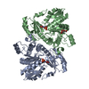
| |||||||||||||||
| Unit cell |
| |||||||||||||||
| Components on special symmetry positions |
|
-Components
| #1: Protein | Mass: 44102.242 Da / Num. of mol.: 2 / Mutation: V244I, L360P Source method: isolated from a genetically manipulated source Details: Covalent linkage between LYS 210 NZ atom and PLP C4A and PLP adduct formed with oxamate. Present in a 10/90 ratio. Source: (gene. exp.) Klebsiella aerogenes KCTC 2190 (bacteria)Gene: EAE_03480 / Plasmid: KlaeA.00906.a.1 / Production host: #2: Chemical |   Mass: 320.193 Da / Num. of mol.: 2 / Source method: obtained synthetically / Formula: C10H13N2O8P / Feature type: SUBJECT OF INVESTIGATION Mass: 320.193 Da / Num. of mol.: 2 / Source method: obtained synthetically / Formula: C10H13N2O8P / Feature type: SUBJECT OF INVESTIGATION#3: Chemical |   Mass: 247.142 Da / Num. of mol.: 2 / Source method: obtained synthetically / Formula: C8H10NO6P / Feature type: SUBJECT OF INVESTIGATION Mass: 247.142 Da / Num. of mol.: 2 / Source method: obtained synthetically / Formula: C8H10NO6P / Feature type: SUBJECT OF INVESTIGATION#4: Chemical | ChemComp-FLC / |   Mass: 189.100 Da / Num. of mol.: 1 / Source method: obtained synthetically / Formula: C6H5O7 Mass: 189.100 Da / Num. of mol.: 1 / Source method: obtained synthetically / Formula: C6H5O7#5: Water | ChemComp-HOH / |  Mass: 18.015 Da / Num. of mol.: 988 / Source method: isolated from a natural source / Formula: H2O Mass: 18.015 Da / Num. of mol.: 988 / Source method: isolated from a natural source / Formula: H2OHas ligand of interest | Y | |
|---|
-Experimental details
-Experiment
| Experiment | Method: X-RAY DIFFRACTION / Number of used crystals: 1 |
|---|
- Sample preparation
| Crystal | Density Matthews: 2.61 Å3/Da / Density % sol: 52.87 % |
|---|---|
| Crystal grow | Temperature: 291 K / Method: vapor diffusion, sitting drop / pH: 8.5 Details: Morpueus G9: 20%(v/v) PEG 500 MME, 10%(w/v) PEG 20000, 100 mM Tris/BICINE, pH 8.5, 20 mM Sodium formate, 20 mM Ammonium acetate, 20 mM Sodium citrate tribasic, 20 mM Potassium sodium ...Details: Morpueus G9: 20%(v/v) PEG 500 MME, 10%(w/v) PEG 20000, 100 mM Tris/BICINE, pH 8.5, 20 mM Sodium formate, 20 mM Ammonium acetate, 20 mM Sodium citrate tribasic, 20 mM Potassium sodium tartrate and 20 mM Sodium oxamate, 2mM PLP added to the protein prior to crystallization, KlaeA.00906.a.B1.PW39169 at 41.1 mg/mL. Plate: 13220, well G9 drop 3, Puck: PSL-0416, Cryo: Direct |
-Data collection
| Diffraction | Mean temperature: 100 K / Serial crystal experiment: N |
|---|---|
| Diffraction source | Source: SYNCHROTRON / Site: NSLS-II / Beamline: 19-ID / Wavelength: 0.9795 Å |
| Detector | Type: DECTRIS EIGER2 XE 9M / Detector: PIXEL / Date: Feb 14, 2022 |
| Radiation | Monochromator: Double Crystal Si 111 / Protocol: SINGLE WAVELENGTH / Monochromatic (M) / Laue (L): M / Scattering type: x-ray |
| Radiation wavelength | Wavelength: 0.9795 Å / Relative weight: 1 |
| Reflection | Resolution: 1.1→80.2 Å / Num. obs: 345136 / % possible obs: 94.7 % / Redundancy: 6.4 % / CC1/2: 0.999 / Rmerge(I) obs: 0.056 / Rpim(I) all: 0.023 / Rrim(I) all: 0.061 / Χ2: 1.02 / Net I/σ(I): 12.4 / Num. measured all: 2207969 |
| Reflection shell | Resolution: 1.1→1.13 Å / % possible obs: 58.7 % / Redundancy: 3.7 % / Rmerge(I) obs: 1.056 / Num. measured all: 59166 / Num. unique obs: 15798 / CC1/2: 0.574 / Rpim(I) all: 0.617 / Rrim(I) all: 1.229 / Χ2: 1.03 / Net I/σ(I) obs: 1.1 |
- Processing
| Software |
| |||||||||||||||||||||||||||||||||||||||||||||||||||||||||||||||||||||||||||||||||||||||||||||||||||||||||||||||||||||||||||||||||||||||||||||||||||||||||||||||||||||||||||||||||||||||||||||||||||||||||||||||||||||||||
|---|---|---|---|---|---|---|---|---|---|---|---|---|---|---|---|---|---|---|---|---|---|---|---|---|---|---|---|---|---|---|---|---|---|---|---|---|---|---|---|---|---|---|---|---|---|---|---|---|---|---|---|---|---|---|---|---|---|---|---|---|---|---|---|---|---|---|---|---|---|---|---|---|---|---|---|---|---|---|---|---|---|---|---|---|---|---|---|---|---|---|---|---|---|---|---|---|---|---|---|---|---|---|---|---|---|---|---|---|---|---|---|---|---|---|---|---|---|---|---|---|---|---|---|---|---|---|---|---|---|---|---|---|---|---|---|---|---|---|---|---|---|---|---|---|---|---|---|---|---|---|---|---|---|---|---|---|---|---|---|---|---|---|---|---|---|---|---|---|---|---|---|---|---|---|---|---|---|---|---|---|---|---|---|---|---|---|---|---|---|---|---|---|---|---|---|---|---|---|---|---|---|---|---|---|---|---|---|---|---|---|---|---|---|---|---|---|---|---|
| Refinement | Method to determine structure: MOLECULAR REPLACEMENT / Resolution: 1.1→26.86 Å / SU ML: 0.08 / Cross valid method: FREE R-VALUE / σ(F): 1.35 / Phase error: 14.59 / Stereochemistry target values: ML
| |||||||||||||||||||||||||||||||||||||||||||||||||||||||||||||||||||||||||||||||||||||||||||||||||||||||||||||||||||||||||||||||||||||||||||||||||||||||||||||||||||||||||||||||||||||||||||||||||||||||||||||||||||||||||
| Solvent computation | Shrinkage radii: 0.9 Å / VDW probe radii: 1.1 Å / Solvent model: FLAT BULK SOLVENT MODEL | |||||||||||||||||||||||||||||||||||||||||||||||||||||||||||||||||||||||||||||||||||||||||||||||||||||||||||||||||||||||||||||||||||||||||||||||||||||||||||||||||||||||||||||||||||||||||||||||||||||||||||||||||||||||||
| Refinement step | Cycle: LAST / Resolution: 1.1→26.86 Å
| |||||||||||||||||||||||||||||||||||||||||||||||||||||||||||||||||||||||||||||||||||||||||||||||||||||||||||||||||||||||||||||||||||||||||||||||||||||||||||||||||||||||||||||||||||||||||||||||||||||||||||||||||||||||||
| Refine LS restraints |
| |||||||||||||||||||||||||||||||||||||||||||||||||||||||||||||||||||||||||||||||||||||||||||||||||||||||||||||||||||||||||||||||||||||||||||||||||||||||||||||||||||||||||||||||||||||||||||||||||||||||||||||||||||||||||
| LS refinement shell |
|
