 Movie
Movie Controller

[English] 日本語
 Yorodumi
- PDB-8s7l: Crystal structure of a double mutant of VirB8-like OrfG central a... -
+ Open data

- Basic information
| Entry | Database: PDB / ID: 8s7l | ||||||
|---|---|---|---|---|---|---|---|
| Title | Crystal structure of a double mutant of VirB8-like OrfG central and C-terminal domains of Streptococcus thermophilus ICESt3 (Gram positive conjugative type IV secretion system). | ||||||
 Components Components | Putative transfer protein | ||||||
 Keywords Keywords | TRANSPORT PROTEIN / VirB8 / Gram-positive / T4SS | ||||||
| Function / homology | Conjugative transposon protein TcpC / TcpC, C-terminal / Conjugative transposon protein TcpC / membrane / Putative transfer protein Function and homology information Function and homology information | ||||||
| Biological species |  Streptococcus thermophilus (bacteria) Streptococcus thermophilus (bacteria) | ||||||
| Method |  X-RAY DIFFRACTION / SYNCHROTRON / MOLECULAR REPLACEMENT / Resolution: 2.6 Å X-RAY DIFFRACTION / SYNCHROTRON / MOLECULAR REPLACEMENT / Resolution: 2.6 Å | ||||||
 Authors Authors | Favier, F. / Didierjean, C. / Maffo-Woulefack, R. / Douzi, B. / Leblond-Bourget, N. | ||||||
| Funding support | 1items
| ||||||
 Citation Citation | Journal: Sci Adv / Year: 2025 Title: Elucidating assembly and function of VirB8 cell wall subunits refines the DNA translocation model in Gram-positive T4SSs. Authors: Maffo-Woulefack, R. / Ali, A.M. / Laroussi, H. / Cappele, J. / Romero-Saavedra, F. / Ramia, N. / Robert, E. / Mathiot, S. / Soler, N. / Roussel, Y. / Fronzes, R. / Huebner, J. / Didierjean, ...Authors: Maffo-Woulefack, R. / Ali, A.M. / Laroussi, H. / Cappele, J. / Romero-Saavedra, F. / Ramia, N. / Robert, E. / Mathiot, S. / Soler, N. / Roussel, Y. / Fronzes, R. / Huebner, J. / Didierjean, C. / Favier, F. / Leblond-Bourget, N. / Douzi, B. | ||||||
| History |
|
- Structure visualization
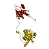
| Structure viewer | Molecule: MolmilJmol/JSmol |
|---|
- Downloads & links
-Download
| PDBx/mmCIF format | 8s7l.cif.gz | 284.7 KB | Display | PDBx/mmCIF format |
|---|---|---|---|---|
| PDB format | pdb8s7l.ent.gz | 229.6 KB | Display | PDB format |
| PDBx/mmJSON format | 8s7l.json.gz | Tree view | PDBx/mmJSON format | |
| Others |  Other downloads Other downloads |
-Validation report
| Summary document | 8s7l_validation.pdf.gz | 451.1 KB | Display | wwPDB validaton report |
|---|---|---|---|---|
| Full document | 8s7l_full_validation.pdf.gz | 465.5 KB | Display | |
| Data in XML | 8s7l_validation.xml.gz | 30.3 KB | Display | |
| Data in CIF | 8s7l_validation.cif.gz | 38.5 KB | Display | |
| Arichive directory | https://data.pdbj.org/pub/pdb/validation_reports/s7/8s7lftp://data.pdbj.org/pub/pdb/validation_reports/s7/8s7l | HTTPS FTP |
-Related structure data
-Links
 PDBj
PDBj- Assembly
| Deposited unit | 
| ||||||||
|---|---|---|---|---|---|---|---|---|---|
| 1 |
| ||||||||
| Unit cell |
|
-Components
| #1: Protein | Mass: 30948.336 Da / Num. of mol.: 3 Source method: isolated from a genetically manipulated source Source: (gene. exp.) Streptococcus thermophilus (bacteria) / Plasmid: PLYSS / Production host: Has protein modification | Y | |
|---|
-Experimental details
-Experiment
| Experiment | Method: X-RAY DIFFRACTION / Number of used crystals: 1 |
|---|
- Sample preparation
| Crystal | Density Matthews: 3.35 Å3/Da / Density % sol: 63.3 % |
|---|---|
| Crystal grow | Temperature: 293 K / Method: vapor diffusion, sitting drop Details: Droplet: 0.3 ul [protein (33mg/ml) in 50 mM Tris pH 8.0 + 100 mM NaCl] + 0.3 ul [reservoir] Reservoir: 50 ul [40% v/v PEG 300, 100 mM potassium phosphate citrate, pH 4.2] |
-Data collection
| Diffraction | Mean temperature: 100 K / Serial crystal experiment: N |
|---|---|
| Diffraction source | Source: SYNCHROTRON / Site: ESRF  / Beamline: MASSIF-3 / Wavelength: 0.967697 Å / Beamline: MASSIF-3 / Wavelength: 0.967697 Å |
| Detector | Type: DECTRIS EIGER X 4M / Detector: PIXEL / Date: Nov 11, 2022 |
| Radiation | Protocol: SINGLE WAVELENGTH / Monochromatic (M) / Laue (L): M / Scattering type: x-ray |
| Radiation wavelength | Wavelength: 0.967697 Å / Relative weight: 1 |
| Reflection | Resolution: 2.6→91.054 Å / Num. obs: 37215 / % possible obs: 98.8 % / Redundancy: 3.1 % / Biso Wilson estimate: 50.28 Å2 / CC1/2: 0.995 / Rmerge(I) obs: 0.062 / Rpim(I) all: 0.061 / Rrim(I) all: 0.087 / Net I/σ(I): 10.6 |
| Reflection shell | Resolution: 2.6→2.72 Å / Redundancy: 3.3 % / Rmerge(I) obs: 0.665 / Mean I/σ(I) obs: 1.8 / Num. unique obs: 4577 / CC1/2: 0.682 / Rpim(I) all: 0.653 / Rrim(I) all: 0.933 / % possible all: 99.8 |
- Processing
| Software |
| |||||||||||||||||||||||||||||||||||||||||||||||||||||||||||||||||||||||||||||||||||||||||||||||||||||||||||||||||||||||||||||||||||||||||||||||||||||||||||||||||||||||||||||||||||||||||||||||||||||||||||||||||||||||||||||||||||||||
|---|---|---|---|---|---|---|---|---|---|---|---|---|---|---|---|---|---|---|---|---|---|---|---|---|---|---|---|---|---|---|---|---|---|---|---|---|---|---|---|---|---|---|---|---|---|---|---|---|---|---|---|---|---|---|---|---|---|---|---|---|---|---|---|---|---|---|---|---|---|---|---|---|---|---|---|---|---|---|---|---|---|---|---|---|---|---|---|---|---|---|---|---|---|---|---|---|---|---|---|---|---|---|---|---|---|---|---|---|---|---|---|---|---|---|---|---|---|---|---|---|---|---|---|---|---|---|---|---|---|---|---|---|---|---|---|---|---|---|---|---|---|---|---|---|---|---|---|---|---|---|---|---|---|---|---|---|---|---|---|---|---|---|---|---|---|---|---|---|---|---|---|---|---|---|---|---|---|---|---|---|---|---|---|---|---|---|---|---|---|---|---|---|---|---|---|---|---|---|---|---|---|---|---|---|---|---|---|---|---|---|---|---|---|---|---|---|---|---|---|---|---|---|---|---|---|---|---|---|---|---|---|---|
| Refinement | Method to determine structure: MOLECULAR REPLACEMENT / Resolution: 2.6→91.054 Å / Cor.coef. Fo:Fc: 0.948 / Cor.coef. Fo:Fc free: 0.921 / SU B: 11.501 / SU ML: 0.231 / Cross valid method: FREE R-VALUE / ESU R: 0.38 / ESU R Free: 0.278 Details: Hydrogens have been added in their riding positions
| |||||||||||||||||||||||||||||||||||||||||||||||||||||||||||||||||||||||||||||||||||||||||||||||||||||||||||||||||||||||||||||||||||||||||||||||||||||||||||||||||||||||||||||||||||||||||||||||||||||||||||||||||||||||||||||||||||||||
| Solvent computation | Ion probe radii: 0.8 Å / Shrinkage radii: 0.8 Å / VDW probe radii: 1.2 Å / Solvent model: BABINET MODEL PLUS MASK | |||||||||||||||||||||||||||||||||||||||||||||||||||||||||||||||||||||||||||||||||||||||||||||||||||||||||||||||||||||||||||||||||||||||||||||||||||||||||||||||||||||||||||||||||||||||||||||||||||||||||||||||||||||||||||||||||||||||
| Displacement parameters | Biso mean: 86.279 Å2
| |||||||||||||||||||||||||||||||||||||||||||||||||||||||||||||||||||||||||||||||||||||||||||||||||||||||||||||||||||||||||||||||||||||||||||||||||||||||||||||||||||||||||||||||||||||||||||||||||||||||||||||||||||||||||||||||||||||||
| Refinement step | Cycle: LAST / Resolution: 2.6→91.054 Å
| |||||||||||||||||||||||||||||||||||||||||||||||||||||||||||||||||||||||||||||||||||||||||||||||||||||||||||||||||||||||||||||||||||||||||||||||||||||||||||||||||||||||||||||||||||||||||||||||||||||||||||||||||||||||||||||||||||||||
| Refine LS restraints |
| |||||||||||||||||||||||||||||||||||||||||||||||||||||||||||||||||||||||||||||||||||||||||||||||||||||||||||||||||||||||||||||||||||||||||||||||||||||||||||||||||||||||||||||||||||||||||||||||||||||||||||||||||||||||||||||||||||||||
| LS refinement shell | Refine-ID: X-RAY DIFFRACTION / Total num. of bins used: 20
|
