 Movie
Movie Controller

[English] 日本語
 Yorodumi
- PDB-8s7i: Fructose 6-phosphate aldolase, L107C/A129G/R134V/L163C/S166G mutant -
+ Open data

- Basic information
| Entry | Database: PDB / ID: 8s7i | ||||||||||||||||||||||||
|---|---|---|---|---|---|---|---|---|---|---|---|---|---|---|---|---|---|---|---|---|---|---|---|---|---|
| Title | Fructose 6-phosphate aldolase, L107C/A129G/R134V/L163C/S166G mutant | ||||||||||||||||||||||||
 Components Components | Fructose-6-phosphate aldolase 1 | ||||||||||||||||||||||||
 Keywords Keywords | LYASE / fructose 6-phosphate aldolase / mutant / carboligation | ||||||||||||||||||||||||
| Function / homology |  Function and homology information Function and homology informationketone catabolic process / fructose 6-phosphate aldolase activity / Lyases; Carbon-carbon lyases; Aldehyde-lyases / fructose metabolic process / identical protein binding / cytoplasm Similarity search - Function | ||||||||||||||||||||||||
| Biological species |  | ||||||||||||||||||||||||
| Method | ELECTRON MICROSCOPY / single particle reconstruction / cryo EM / Resolution: 3.2 Å | ||||||||||||||||||||||||
 Authors Authors | Hebert, H. / Widersten, M. | ||||||||||||||||||||||||
| Funding support |  Sweden, 1items Sweden, 1items
| ||||||||||||||||||||||||
 Citation Citation | Journal: Structure / Year: 2024 Title: Structure of the iminium reaction intermediate in an engineered aldolase explains the carboligation activity toward arylated ketones and aldehydes. Authors: Hans Hebert / Eda Sönmez / Pasi Purhonen / Mikael Widersten / Abstract: Two structures of fructose 6-phosphate aldolase, the wild-type and an engineered variant containing five active-site mutations, have been solved by cryoelectron microscopy (cryo-EM). The engineered ...Two structures of fructose 6-phosphate aldolase, the wild-type and an engineered variant containing five active-site mutations, have been solved by cryoelectron microscopy (cryo-EM). The engineered variant affords production of aldols from aryl substituted ketones and aldehydes. This structure was solved to a resolution of 3.1 Å and contains the critical iminium reaction intermediate trapped in the active site. This provides new information that rationalizes the acquired substrate scope and aids in formulating hypotheses of the chemical mechanism. A Tyr residue (Y131) is positioned for a role as catalytic acid/base during the aldol reaction and the different structures demonstrate mobility of this amino acid residue. Further engineering of this fructose 6-phosphate aldolase (FSA) variant, guided by this new structure, identified additional FSA variants that display improved carboligation activities with 2-hydroxyacetophenone and phenylacetaldehyde. | ||||||||||||||||||||||||
| History |
|
- Structure visualization
| Structure viewer | Molecule: MolmilJmol/JSmol |
|---|
- Downloads & links
-Download
| PDBx/mmCIF format | 8s7i.cif.gz | 759.5 KB | Display | PDBx/mmCIF format |
|---|---|---|---|---|
| PDB format | pdb8s7i.ent.gz | Display | PDB format | |
| PDBx/mmJSON format | 8s7i.json.gz | Tree view | PDBx/mmJSON format | |
| Others |  Other downloads Other downloads |
-Validation report
| Arichive directory | https://data.pdbj.org/pub/pdb/validation_reports/s7/8s7iftp://data.pdbj.org/pub/pdb/validation_reports/s7/8s7i | HTTPS FTP |
|---|
-Related structure data
| Related structure data |  19773MC 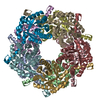 8s7hC M: map data used to model this data C: citing same article ( |
|---|---|
| Similar structure data |

-Links
 PDBj
PDBj- Assembly
| Deposited unit | 
|
|---|---|
| 1 |
|
-Components
| #1: Protein | Mass: 23890.650 Da / Num. of mol.: 10 Source method: isolated from a genetically manipulated source Details: The TSHHHHH is a C-terminal His-tag not observed in the map Source: (gene. exp.) References: UniProt: P78055, Lyases; Carbon-carbon lyases; Aldehyde-lyases #2: Water | ChemComp-HOH / |  Mass: 18.015 Da / Num. of mol.: 272 / Source method: isolated from a natural source / Formula: H2O Mass: 18.015 Da / Num. of mol.: 272 / Source method: isolated from a natural source / Formula: H2OHas ligand of interest | Y | Has protein modification | Y | |
|---|
-Experimental details
-Experiment
| Experiment | Method: ELECTRON MICROSCOPY |
|---|---|
| EM experiment | Aggregation state: PARTICLE / 3D reconstruction method: single particle reconstruction |
- Sample preparation
| Component | Name: Fructose 6-phosphate aldolase, L107C/A129G/R134V/L163C/S166G mutant Type: COMPLEX / Entity ID: #1 / Source: RECOMBINANT |
|---|---|
| Source (natural) | Organism: |
| Source (recombinant) | Organism: |
| Buffer solution | pH: 8 |
| Buffer component | Conc.: 40 mM / Name: bicine / Formula: C6H13NO4 |
| Specimen | Conc.: 2 mg/ml / Embedding applied: NO / Shadowing applied: NO / Staining applied: NO / Vitrification applied: YES |
| Specimen support | Grid material: COPPER / Grid mesh size: 200 divisions/in. / Grid type: Quantifoil R2/1 |
| Vitrification | Instrument: FEI VITROBOT MARK I / Cryogen name: ETHANE / Humidity: 100 % / Chamber temperature: 289 K |
- Electron microscopy imaging
| Experimental equipment |  Model: Titan Krios / Image courtesy: FEI Company |
|---|---|
| Microscopy | Model: FEI TITAN KRIOS |
| Electron gun | Electron source:  FIELD EMISSION GUN / Accelerating voltage: 300 kV / Illumination mode: FLOOD BEAM FIELD EMISSION GUN / Accelerating voltage: 300 kV / Illumination mode: FLOOD BEAM |
| Electron lens | Mode: BRIGHT FIELD / Nominal magnification: 105000 X / Nominal defocus max: 2000 nm / Nominal defocus min: 600 nm / Cs: 2.7 mm / C2 aperture diameter: 50 µm |
| Specimen holder | Cryogen: NITROGEN / Specimen holder model: FEI TITAN KRIOS AUTOGRID HOLDER |
| Image recording | Average exposure time: 2.2 sec. / Electron dose: 50.04 e/Å2 / Film or detector model: GATAN K3 (6k x 4k) / Num. of grids imaged: 1 / Num. of real images: 8165 Details: Images were collected in movie-mode, 40 frames with 1.251 e-/A2 dose/frame |
| EM imaging optics | Energyfilter slit width: 20 eV |
| Image scans | Width: 5760 / Height: 4092 |
- Processing
| EM software |
| |||||||||||||||||||||||||||
|---|---|---|---|---|---|---|---|---|---|---|---|---|---|---|---|---|---|---|---|---|---|---|---|---|---|---|---|---|
| CTF correction | Type: PHASE FLIPPING ONLY | |||||||||||||||||||||||||||
| Particle selection | Num. of particles selected: 307612 | |||||||||||||||||||||||||||
| Symmetry | Point symmetry: D5 (2x5 fold dihedral) | |||||||||||||||||||||||||||
| 3D reconstruction | Resolution: 3.2 Å / Resolution method: FSC 0.143 CUT-OFF / Num. of particles: 115055 / Num. of class averages: 1 / Symmetry type: POINT | |||||||||||||||||||||||||||
| Atomic model building | B value: 156 / Protocol: FLEXIBLE FIT / Space: REAL Details: Initial local fitting was done using Chimera and Phenix was used for flexible fitting | |||||||||||||||||||||||||||
| Atomic model building | PDB-ID: 3wnq Accession code: 3wnq / Source name: PDB / Type: experimental model | |||||||||||||||||||||||||||
| Refine LS restraints |
|

