 Movie
Movie Controller

[English] 日本語
 Yorodumi
- PDB-8s4m: Crystal structure of Mycobacterium tuberculosis cytochrome P450 C... -
+ Open data

- Basic information
| Entry | Database: PDB / ID: 8s4m | ||||||
|---|---|---|---|---|---|---|---|
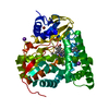
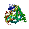
| Title | Crystal structure of Mycobacterium tuberculosis cytochrome P450 CYP125 in complex with an inhibitor | ||||||
 Components Components | Steroid C26-monooxygenase | ||||||
 Keywords Keywords | OXIDOREDUCTASE / CYP125 / tuberculosis / cholesterol / P450 / CYP / cytochrome / fragment / cholestenone | ||||||
| Function / homology |  Function and homology information Function and homology informationcholest-4-en-3-one 26-monooxygenase [(25S)-3-oxocholest-4-en-26-oate forming] / cholest-4-en-3-one 26-monooxygenase activity / biological process involved in interaction with host / steroid hydroxylase activity / cholesterol catabolic process / iron ion binding / heme binding Similarity search - Function | ||||||
| Biological species |  Mycobacterium tuberculosis H37Rv (bacteria) Mycobacterium tuberculosis H37Rv (bacteria) | ||||||
| Method |  X-RAY DIFFRACTION / SYNCHROTRON / MOLECULAR REPLACEMENT / Resolution: 2.1 Å X-RAY DIFFRACTION / SYNCHROTRON / MOLECULAR REPLACEMENT / Resolution: 2.1 Å | ||||||
 Authors Authors | Snee, M. / Kavanagh, M. / Levy, C. | ||||||
| Funding support |  United Kingdom, 1items United Kingdom, 1items
| ||||||
 Citation Citation | Journal: J.Med.Chem. / Year: 2025 Title: Fragment-Based Development of Small Molecule Inhibitors Targeting Mycobacterium tuberculosis Cholesterol Metabolism. Authors: Kavanagh, M.E. / McLean, K.J. / Gilbert, S.H. / Amadi, C.N. / Snee, M. / Tunnicliffe, R.B. / Arora, K. / Boshoff, H.I.M. / Fanourakis, A. / Rebollo-Lopez, M.J. / Ortega, F. / Levy, C.W. / ...Authors: Kavanagh, M.E. / McLean, K.J. / Gilbert, S.H. / Amadi, C.N. / Snee, M. / Tunnicliffe, R.B. / Arora, K. / Boshoff, H.I.M. / Fanourakis, A. / Rebollo-Lopez, M.J. / Ortega, F. / Levy, C.W. / Munro, A.W. / Leys, D. / Abell, C. / Coyne, A.G. #1: Journal: Biorxiv / Year: 2024 Title: Fragment-based development of small molecule inhibitors targeting Mycobacterium tuberculosis cholesterol metabolism. Authors: Kavanagh, M.E. / McLean, K.J. / Gilbert, S.H. / Amadi, C. / Snee, M. / Tunnicliffe, R.B. / Arora, K. / Boshoff, H.I. / Fanourakis, A. / Rebello-Lopez, M.J. / Ortega-Muro, F. / Levy, C.W. / ...Authors: Kavanagh, M.E. / McLean, K.J. / Gilbert, S.H. / Amadi, C. / Snee, M. / Tunnicliffe, R.B. / Arora, K. / Boshoff, H.I. / Fanourakis, A. / Rebello-Lopez, M.J. / Ortega-Muro, F. / Levy, C.W. / Munro, A.W. / Leys, D. / Abell, C. / Coyne, A.G. #2: Journal: Acta Crystallogr., Sect. D: Biol. Crystallogr. / Year: 2012Title: Towards automated crystallographic structure refinement with phenix.refine. Authors: Afonine, P. / Grosse-Kunstleve, R. / Echols, N. | ||||||
| History |
|
- Structure visualization
| Structure viewer | Molecule: MolmilJmol/JSmol |
|---|
- Downloads & links
-Download
| PDBx/mmCIF format | 8s4m.cif.gz | 861.4 KB | Display | PDBx/mmCIF format |
|---|---|---|---|---|
| PDB format | pdb8s4m.ent.gz | Display | PDB format | |
| PDBx/mmJSON format | 8s4m.json.gz | Tree view | PDBx/mmJSON format | |
| Others |  Other downloads Other downloads |
-Validation report
| Arichive directory | https://data.pdbj.org/pub/pdb/validation_reports/s4/8s4mftp://data.pdbj.org/pub/pdb/validation_reports/s4/8s4m | HTTPS FTP |
|---|
-Related structure data

-Links
 PDBj
PDBj
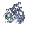
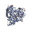
- Assembly
| Deposited unit | 
| ||||||||||||
|---|---|---|---|---|---|---|---|---|---|---|---|---|---|
| 1 | 
| ||||||||||||
| 2 | 
| ||||||||||||
| 3 | 
| ||||||||||||
| Unit cell |
|
-Components
| #1: Protein | Mass: 46663.383 Da / Num. of mol.: 3 Source method: isolated from a genetically manipulated source Source: (gene. exp.) Mycobacterium tuberculosis H37Rv (bacteria)Gene: cyp125, cyp125A1, Rv3545c, MTCY03C7.11 / Production host: References: UniProt: P9WPP1, cholest-4-en-3-one 26-monooxygenase [(25S)-3-oxocholest-4-en-26-oate forming] #2: Chemical |   Mass: 616.487 Da / Num. of mol.: 3 / Source method: obtained synthetically / Formula: C34H32FeN4O4 Mass: 616.487 Da / Num. of mol.: 3 / Source method: obtained synthetically / Formula: C34H32FeN4O4#3: Chemical | ChemComp-SO4 /   Mass: 96.063 Da / Num. of mol.: 10 / Source method: obtained synthetically / Formula: SO4 Mass: 96.063 Da / Num. of mol.: 10 / Source method: obtained synthetically / Formula: SO4#4: Chemical | ChemComp-A1H47 / ~{ | Mass: 342.394 Da / Num. of mol.: 1 / Source method: obtained synthetically / Formula: C21H18N4O / Feature type: SUBJECT OF INVESTIGATION #5: Water | ChemComp-HOH / |  Mass: 18.015 Da / Num. of mol.: 328 / Source method: isolated from a natural source / Formula: H2O Mass: 18.015 Da / Num. of mol.: 328 / Source method: isolated from a natural source / Formula: H2OHas ligand of interest | Y | Has protein modification | N | |
|---|
-Experimental details
-Experiment
| Experiment | Method: X-RAY DIFFRACTION / Number of used crystals: 1 |
|---|
- Sample preparation
| Crystal | Density Matthews: 2.42 Å3/Da / Density % sol: 49.19 % / Description: chunks |
|---|---|
| Crystal grow | Temperature: 277.15 K / Method: vapor diffusion, sitting drop / pH: 6.2 / Details: 1.9 M Ammonium sulphate, 0.1M MES pH 6.2 / PH range: 6-6.5 / Temp details: 4C |
-Data collection
| Diffraction | Mean temperature: 100 K / Ambient temp details: N2 cryostream / Serial crystal experiment: N |
|---|---|
| Diffraction source | Source: SYNCHROTRON / Site: Diamond / Beamline: I04-1 / Wavelength: 0.9159 Å |
| Detector | Type: DECTRIS PILATUS 6M / Detector: PIXEL / Date: Jul 7, 2019 |
| Radiation | Protocol: SINGLE WAVELENGTH / Monochromatic (M) / Laue (L): M / Scattering type: x-ray |
| Radiation wavelength | Wavelength: 0.9159 Å / Relative weight: 1 |
| Reflection | Resolution: 2.1→68.29 Å / Num. obs: 78300 / % possible obs: 99.9 % / Redundancy: 3.3 % / Biso Wilson estimate: 38.76 Å2 / CC1/2: 0.995 / Rmerge(I) obs: 0.071 / Rpim(I) all: 0.046 / Rrim(I) all: 0.084 / Net I/σ(I): 9.1 |
| Reflection shell | Resolution: 2.1→2.14 Å / Rmerge(I) obs: 0.861 / Mean I/σ(I) obs: 1.4 / Num. unique obs: 4463 / CC1/2: 0.585 / Rpim(I) all: 0.545 / Rrim(I) all: 1.022 |
- Processing
| Software |
| |||||||||||||||||||||||||||||||||||||||||||||||||||||||||||||||||||||||||||||||||||||||||||||||||||||||||||||||||||||||||||||||||||||||||||||||||||||||||||||||||||||||||||||||||||||||||||||||||||||||||||
|---|---|---|---|---|---|---|---|---|---|---|---|---|---|---|---|---|---|---|---|---|---|---|---|---|---|---|---|---|---|---|---|---|---|---|---|---|---|---|---|---|---|---|---|---|---|---|---|---|---|---|---|---|---|---|---|---|---|---|---|---|---|---|---|---|---|---|---|---|---|---|---|---|---|---|---|---|---|---|---|---|---|---|---|---|---|---|---|---|---|---|---|---|---|---|---|---|---|---|---|---|---|---|---|---|---|---|---|---|---|---|---|---|---|---|---|---|---|---|---|---|---|---|---|---|---|---|---|---|---|---|---|---|---|---|---|---|---|---|---|---|---|---|---|---|---|---|---|---|---|---|---|---|---|---|---|---|---|---|---|---|---|---|---|---|---|---|---|---|---|---|---|---|---|---|---|---|---|---|---|---|---|---|---|---|---|---|---|---|---|---|---|---|---|---|---|---|---|---|---|---|---|---|---|---|
| Refinement | Method to determine structure: MOLECULAR REPLACEMENT / Resolution: 2.1→68.29 Å / SU ML: 0.2739 / Cross valid method: FREE R-VALUE / σ(F): 1.34 / Phase error: 27.2907 Stereochemistry target values: GeoStd + Monomer Library + CDL v1.2
| |||||||||||||||||||||||||||||||||||||||||||||||||||||||||||||||||||||||||||||||||||||||||||||||||||||||||||||||||||||||||||||||||||||||||||||||||||||||||||||||||||||||||||||||||||||||||||||||||||||||||||
| Solvent computation | Shrinkage radii: 0.9 Å / VDW probe radii: 1.11 Å / Solvent model: FLAT BULK SOLVENT MODEL | |||||||||||||||||||||||||||||||||||||||||||||||||||||||||||||||||||||||||||||||||||||||||||||||||||||||||||||||||||||||||||||||||||||||||||||||||||||||||||||||||||||||||||||||||||||||||||||||||||||||||||
| Displacement parameters | Biso mean: 59.24 Å2 | |||||||||||||||||||||||||||||||||||||||||||||||||||||||||||||||||||||||||||||||||||||||||||||||||||||||||||||||||||||||||||||||||||||||||||||||||||||||||||||||||||||||||||||||||||||||||||||||||||||||||||
| Refinement step | Cycle: LAST / Resolution: 2.1→68.29 Å
| |||||||||||||||||||||||||||||||||||||||||||||||||||||||||||||||||||||||||||||||||||||||||||||||||||||||||||||||||||||||||||||||||||||||||||||||||||||||||||||||||||||||||||||||||||||||||||||||||||||||||||
| Refine LS restraints |
| |||||||||||||||||||||||||||||||||||||||||||||||||||||||||||||||||||||||||||||||||||||||||||||||||||||||||||||||||||||||||||||||||||||||||||||||||||||||||||||||||||||||||||||||||||||||||||||||||||||||||||
| LS refinement shell |
| |||||||||||||||||||||||||||||||||||||||||||||||||||||||||||||||||||||||||||||||||||||||||||||||||||||||||||||||||||||||||||||||||||||||||||||||||||||||||||||||||||||||||||||||||||||||||||||||||||||||||||
| Refinement TLS params. | Method: refined / Origin x: 29.5723410479 Å / Origin y: -4.46821048012 Å / Origin z: 37.7477922759 Å
| |||||||||||||||||||||||||||||||||||||||||||||||||||||||||||||||||||||||||||||||||||||||||||||||||||||||||||||||||||||||||||||||||||||||||||||||||||||||||||||||||||||||||||||||||||||||||||||||||||||||||||
| Refinement TLS group | Selection details: all |
