 Movie
Movie Controller

[English] 日本語
 Yorodumi
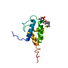
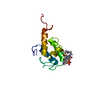
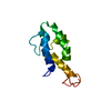
- PDB-8rz6: SeMet derivative structure of the condensation domain TomBC from ... -
+ Open data

- Basic information
| Entry | Database: PDB / ID: 8rz6 | ||||||
|---|---|---|---|---|---|---|---|
| Title | SeMet derivative structure of the condensation domain TomBC from the Tomaymycin non-ribosomal peptide synthetase | ||||||
 Components Components | Tomaymycin non-ribosomal peptide synthetase | ||||||
 Keywords Keywords | BIOSYNTHETIC PROTEIN / Non-ribosomal peptide synthetase / Tomaymycin / Condensation | ||||||
| Function / homology | FORMIC ACID / :  Function and homology information Function and homology information | ||||||
| Biological species |  Streptomyces regensis (bacteria) Streptomyces regensis (bacteria) | ||||||
| Method |  X-RAY DIFFRACTION / SYNCHROTRON / SAD / Resolution: 2.3 Å X-RAY DIFFRACTION / SYNCHROTRON / SAD / Resolution: 2.3 Å | ||||||
 Authors Authors | Karanth, M. / Schmelz, S. / Kirkpatrick, J. / Krausze, J. / Scrima, A. / Carlomagno, T. | ||||||
| Funding support |  Germany, 1items Germany, 1items
| ||||||
 Citation Citation | Journal: Sci Adv / Year: 2024 Title: The specificity of intermodular recognition in a prototypical nonribosomal peptide synthetase depends on an adaptor domain. Authors: Karanth, M.N. / Kirkpatrick, J.P. / Krausze, J. / Schmelz, S. / Scrima, A. / Carlomagno, T. | ||||||
| History |
|
- Structure visualization
| Structure viewer | Molecule: MolmilJmol/JSmol |
|---|
- Downloads & links
-Download
| PDBx/mmCIF format | 8rz6.cif.gz | 348.6 KB | Display | PDBx/mmCIF format |
|---|---|---|---|---|
| PDB format | pdb8rz6.ent.gz | 285.5 KB | Display | PDB format |
| PDBx/mmJSON format | 8rz6.json.gz | Tree view | PDBx/mmJSON format | |
| Others |  Other downloads Other downloads |
-Validation report
| Arichive directory | https://data.pdbj.org/pub/pdb/validation_reports/rz/8rz6ftp://data.pdbj.org/pub/pdb/validation_reports/rz/8rz6 | HTTPS FTP |
|---|
-Related structure data

-Links
 PDBj
PDBj- Assembly
| Deposited unit | 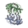
| ||||||||
|---|---|---|---|---|---|---|---|---|---|
| 1 |
| ||||||||
| Unit cell |
|
-Components
| #1: Protein | Mass: 59713.070 Da / Num. of mol.: 2 Source method: isolated from a genetically manipulated source Details: SeMet derivative structure of the condensation domain TomBC from Tomaymycin non-ribosomal peptide synthetase Source: (gene. exp.) Streptomyces regensis (bacteria) / Variant: FH6421 / Production host: #2: Chemical | ChemComp-FMT /   Mass: 46.025 Da / Num. of mol.: 4 / Source method: obtained synthetically / Formula: CH2O2 Mass: 46.025 Da / Num. of mol.: 4 / Source method: obtained synthetically / Formula: CH2O2#3: Chemical | ChemComp-K /   Mass: 39.098 Da / Num. of mol.: 4 / Source method: obtained synthetically / Formula: K Mass: 39.098 Da / Num. of mol.: 4 / Source method: obtained synthetically / Formula: K#4: Chemical |   Mass: 92.094 Da / Num. of mol.: 3 / Source method: obtained synthetically / Formula: C3H8O3 Mass: 92.094 Da / Num. of mol.: 3 / Source method: obtained synthetically / Formula: C3H8O3#5: Water | ChemComp-HOH / |  Mass: 18.015 Da / Num. of mol.: 586 / Source method: isolated from a natural source / Formula: H2O Mass: 18.015 Da / Num. of mol.: 586 / Source method: isolated from a natural source / Formula: H2OHas ligand of interest | N | Has protein modification | Y | |
|---|
-Experimental details
-Experiment
| Experiment | Method: X-RAY DIFFRACTION / Number of used crystals: 1 |
|---|
- Sample preparation
| Crystal | Density Matthews: 3.43 Å3/Da / Density % sol: 64.11 % |
|---|---|
| Crystal grow | Temperature: 277 K / Method: vapor diffusion, sitting drop Details: 100 mM Tris pH 8.5, 3.0 M KFormate, with 25% glycerol used as cryoprotectant |
-Data collection
| Diffraction | Mean temperature: 100 K / Serial crystal experiment: N |
|---|---|
| Diffraction source | Source: SYNCHROTRON / Site: BESSY / Beamline: 14.1 / Wavelength: 0.979733 Å |
| Detector | Type: DECTRIS PILATUS 6M / Detector: PIXEL / Date: Jul 7, 2015 |
| Radiation | Protocol: SINGLE WAVELENGTH / Monochromatic (M) / Laue (L): M / Scattering type: x-ray |
| Radiation wavelength | Wavelength: 0.979733 Å / Relative weight: 1 |
| Reflection | Resolution: 2.3→48.91 Å / Num. obs: 139112 / % possible obs: 99.1 % / Redundancy: 10.58 % / CC1/2: 0.994 / Net I/σ(I): 9.9 |
| Reflection shell | Resolution: 2.3→2.4 Å / Redundancy: 10.82 % / Mean I/σ(I) obs: 2.1 / Num. unique obs: 16569 / CC1/2: 0.747 / % possible all: 98.7 |
- Processing
| Software |
| ||||||||||||||||||||||||||||||||||||||||||||||||||||||||||||||||||||||||||||||||||||||||||||||||||||||||||||||||||||||||||||||||||||||||||||||||||||||||||||||||||||||||||||||||||||||
|---|---|---|---|---|---|---|---|---|---|---|---|---|---|---|---|---|---|---|---|---|---|---|---|---|---|---|---|---|---|---|---|---|---|---|---|---|---|---|---|---|---|---|---|---|---|---|---|---|---|---|---|---|---|---|---|---|---|---|---|---|---|---|---|---|---|---|---|---|---|---|---|---|---|---|---|---|---|---|---|---|---|---|---|---|---|---|---|---|---|---|---|---|---|---|---|---|---|---|---|---|---|---|---|---|---|---|---|---|---|---|---|---|---|---|---|---|---|---|---|---|---|---|---|---|---|---|---|---|---|---|---|---|---|---|---|---|---|---|---|---|---|---|---|---|---|---|---|---|---|---|---|---|---|---|---|---|---|---|---|---|---|---|---|---|---|---|---|---|---|---|---|---|---|---|---|---|---|---|---|---|---|---|---|
| Refinement | Method to determine structure: SAD / Resolution: 2.3→48.91 Å / SU ML: 0.27 / Cross valid method: THROUGHOUT / σ(F): 1.36 / Phase error: 21.75 / Stereochemistry target values: ML
| ||||||||||||||||||||||||||||||||||||||||||||||||||||||||||||||||||||||||||||||||||||||||||||||||||||||||||||||||||||||||||||||||||||||||||||||||||||||||||||||||||||||||||||||||||||||
| Solvent computation | Shrinkage radii: 0.9 Å / VDW probe radii: 1.11 Å / Solvent model: FLAT BULK SOLVENT MODEL | ||||||||||||||||||||||||||||||||||||||||||||||||||||||||||||||||||||||||||||||||||||||||||||||||||||||||||||||||||||||||||||||||||||||||||||||||||||||||||||||||||||||||||||||||||||||
| Refinement step | Cycle: LAST / Resolution: 2.3→48.91 Å
| ||||||||||||||||||||||||||||||||||||||||||||||||||||||||||||||||||||||||||||||||||||||||||||||||||||||||||||||||||||||||||||||||||||||||||||||||||||||||||||||||||||||||||||||||||||||
| Refine LS restraints |
| ||||||||||||||||||||||||||||||||||||||||||||||||||||||||||||||||||||||||||||||||||||||||||||||||||||||||||||||||||||||||||||||||||||||||||||||||||||||||||||||||||||||||||||||||||||||
| LS refinement shell |
|
