 Movie
Movie Controller


+ Open data

- Basic information
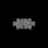
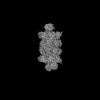
| Entry | Database: PDB / ID: 8rvq | ||||||||||||
|---|---|---|---|---|---|---|---|---|---|---|---|---|---|
| Title | 20S proteasome from pre1-1 | ||||||||||||
 Components Components |
| ||||||||||||
 Keywords Keywords | HYDROLASE / proteasome biogenesis / Ump1 / pre1-1 / cryo-EM / propertied maturation | ||||||||||||
| Function / homology |  Function and homology information Function and homology informationER-Phagosome pathway / Antigen processing: Ub, ATP-independent proteasomal degradation / proteasome core complex assembly / Proteasome assembly / Cross-presentation of soluble exogenous antigens (endosomes) / TNFR2 non-canonical NF-kB pathway / nuclear outer membrane-endoplasmic reticulum membrane network / Regulation of PTEN stability and activity / CDK-mediated phosphorylation and removal of Cdc6 / FBXL7 down-regulates AURKA during mitotic entry and in early mitosis ...ER-Phagosome pathway / Antigen processing: Ub, ATP-independent proteasomal degradation / proteasome core complex assembly / Proteasome assembly / Cross-presentation of soluble exogenous antigens (endosomes) / TNFR2 non-canonical NF-kB pathway / nuclear outer membrane-endoplasmic reticulum membrane network / Regulation of PTEN stability and activity / CDK-mediated phosphorylation and removal of Cdc6 / FBXL7 down-regulates AURKA during mitotic entry and in early mitosis / KEAP1-NFE2L2 pathway / Neddylation / Ubiquitin-Mediated Degradation of Phosphorylated Cdc25A / Orc1 removal from chromatin / MAPK6/MAPK4 signaling / Antigen processing: Ubiquitination & Proteasome degradation / proteasome storage granule / proteasomal ubiquitin-independent protein catabolic process / Ub-specific processing proteases / proteasome endopeptidase complex / proteasome core complex, beta-subunit complex / endopeptidase activator activity / threonine-type endopeptidase activity / proteasome assembly / proteasome core complex, alpha-subunit complex / Neutrophil degranulation / proteasome complex / peroxisome / endopeptidase activity / proteasome-mediated ubiquitin-dependent protein catabolic process / mRNA binding / endoplasmic reticulum membrane / mitochondrion / nucleus / cytosol Similarity search - Function | ||||||||||||
| Biological species |  | ||||||||||||
| Method | ELECTRON MICROSCOPY / single particle reconstruction / cryo EM / Resolution: 2.02 Å | ||||||||||||
 Authors Authors | Mark, E. / Ramos, P.C. / Kayser, F. / Hoeckendorff, J. / Dohmen, R.J. / Wendler, P. | ||||||||||||
| Funding support |  Germany, European Union, 3items Germany, European Union, 3items
| ||||||||||||
 Citation Citation | Journal: Life Sci Alliance / Year: 2024 Title: Structural roles of Ump1 and β-subunit propeptides in proteasome biogenesis. Authors: Eric Mark / Paula C Ramos / Fleur Kayser / Jörg Höckendorff / R Jürgen Dohmen / Petra Wendler / Abstract: The yeast (β4-S142F) mutant accumulates late 20S proteasome core particle precursor complexes (late-PCs). We report a 2.1 Å cryo-EM structure of this intermediate with full-length Ump1 trapped ...The yeast (β4-S142F) mutant accumulates late 20S proteasome core particle precursor complexes (late-PCs). We report a 2.1 Å cryo-EM structure of this intermediate with full-length Ump1 trapped inside, and Pba1-Pba2 attached to the α-ring surfaces. The structure discloses intimate interactions of Ump1 with β2- and β5-propeptides, which together fill most of the antechambers between the α- and β-rings. The β5-propeptide is unprocessed and separates Ump1 from β6 and β7. The β2-propeptide is disconnected from the subunit by autocatalytic processing and localizes between Ump1 and β3. A comparison of different proteasome maturation states reveals that maturation goes along with global conformational changes in the rings, initiated by structuring of the proteolytic sites and their autocatalytic activation. In the strain, β2 is activated first enabling processing of β1-, β6-, and β7-propeptides. Subsequent maturation of β5 and β1 precedes degradation of Ump1, tightening of the complex, and finally release of Pba1-Pba2. | ||||||||||||
| History |
|
- Structure visualization
| Structure viewer | Molecule: MolmilJmol/JSmol |
|---|
- Downloads & links
-Download
| PDBx/mmCIF format | 8rvq.cif.gz | 1.5 MB | Display | PDBx/mmCIF format |
|---|---|---|---|---|
| PDB format | pdb8rvq.ent.gz | 1012.5 KB | Display | PDB format |
| PDBx/mmJSON format | 8rvq.json.gz | Tree view | PDBx/mmJSON format | |
| Others |  Other downloads Other downloads |
-Validation report
| Arichive directory | https://data.pdbj.org/pub/pdb/validation_reports/rv/8rvqftp://data.pdbj.org/pub/pdb/validation_reports/rv/8rvq | HTTPS FTP |
|---|
-Related structure data
| Related structure data | 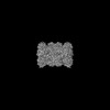 19529MC  8rvlC 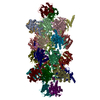 8rvoC  8rvpC  9gbkC M: map data used to model this data C: citing same article ( |
|---|---|
| Similar structure data |


-Links
 PDBj
PDBj


- Assembly
| Deposited unit | 
|
|---|---|
| 1 |
|
-Components
-Proteasome subunit beta type- ... , 7 types, 14 molecules 1M2NHVIWJXLZKY
| #1: Protein | Mass: 24883.928 Da / Num. of mol.: 2 / Source method: isolated from a natural source / Source: (natural) #2: Protein | Mass: 25945.496 Da / Num. of mol.: 2 / Source method: isolated from a natural source / Source: (natural) #6: Protein | Mass: 21517.186 Da / Num. of mol.: 2 / Source method: isolated from a natural source / Source: (natural) References: UniProt: P38624, proteasome endopeptidase complex #7: Protein | Mass: 25114.459 Da / Num. of mol.: 2 / Source method: isolated from a natural source / Source: (natural) References: UniProt: P25043, proteasome endopeptidase complex #8: Protein | Mass: 22627.842 Da / Num. of mol.: 2 / Source method: isolated from a natural source / Source: (natural) #9: Protein | Mass: 23325.248 Da / Num. of mol.: 2 / Source method: isolated from a natural source / Source: (natural) References: UniProt: P30656, proteasome endopeptidase complex #14: Protein | Mass: 24431.629 Da / Num. of mol.: 2 / Mutation: S142F Source method: isolated from a genetically manipulated source Source: (gene. exp.) Gene: PRE1, YER012W / Production host: |
|---|
-Proteasome subunit alpha type- ... , 6 types, 12 molecules DRFTAOBPCQES
| #3: Protein | Mass: 28478.111 Da / Num. of mol.: 2 / Source method: isolated from a natural source / Source: (natural) #4: Protein | Mass: 25634.000 Da / Num. of mol.: 2 / Source method: isolated from a natural source / Source: (natural) #10: Protein | Mass: 28033.830 Da / Num. of mol.: 2 / Source method: isolated from a natural source / Source: (natural) #11: Protein | Mass: 27191.828 Da / Num. of mol.: 2 / Source method: isolated from a natural source / Source: (natural) #12: Protein | Mass: 28748.230 Da / Num. of mol.: 2 / Source method: isolated from a natural source / Source: (natural) #13: Protein | Mass: 28649.086 Da / Num. of mol.: 2 / Source method: isolated from a natural source / Source: (natural) |
|---|
-Protein / Non-polymers , 2 types, 2414 molecules GU
| #15: Water | ChemComp-HOH / Mass: 18.015 Da / Num. of mol.: 2412 / Source method: isolated from a natural source / Formula: H2O |
|---|---|
| #5: Protein | Mass: 31575.068 Da / Num. of mol.: 2 / Source method: isolated from a natural source / Source: (natural) |
-Experimental details
-Experiment
| Experiment | Method: ELECTRON MICROSCOPY |
|---|---|
| EM experiment | Aggregation state: PARTICLE / 3D reconstruction method: single particle reconstruction |
- Sample preparation
| Component | Name: 20S proteasome from pre1-1 / Type: COMPLEX / Entity ID: #1-#14 / Source: NATURAL |
|---|---|
| Molecular weight | Experimental value: NO |
| Source (natural) | Organism: |
| Buffer solution | pH: 7.5 / Details: 150 mM NaCl 50 mM Tris-HCl |
| Specimen | Conc.: 0.35 mg/ml / Embedding applied: NO / Shadowing applied: NO / Staining applied: NO / Vitrification applied: YES |
| Specimen support | Grid material: COPPER / Grid mesh size: 300 divisions/in. / Grid type: Quantifoil R2/4 |
| Vitrification | Instrument: HOMEMADE PLUNGER / Cryogen name: ETHANE / Humidity: 80 % / Chamber temperature: 277 K Details: manual plunge freezing device purchased from 'Neptune Fluid Flow Systems' |
- Electron microscopy imaging
| Experimental equipment |  Model: Titan Krios / Image courtesy: FEI Company |
|---|---|
| Microscopy | Model: FEI TITAN KRIOS |
| Electron gun | Electron source:  FIELD EMISSION GUN / Accelerating voltage: 300 kV / Illumination mode: FLOOD BEAM FIELD EMISSION GUN / Accelerating voltage: 300 kV / Illumination mode: FLOOD BEAM |
| Electron lens | Mode: BRIGHT FIELD / Nominal magnification: 105000 X / Nominal defocus max: 1800 nm / Nominal defocus min: 600 nm / Cs: 2.7 mm |
| Specimen holder | Specimen holder model: FEI TITAN KRIOS AUTOGRID HOLDER |
| Image recording | Average exposure time: 2 sec. / Electron dose: 44 e/Å2 / Film or detector model: GATAN K3 BIOQUANTUM (6k x 4k) / Num. of grids imaged: 1 / Num. of real images: 29904 |
| EM imaging optics | Phase plate: VOLTA PHASE PLATE |
- Processing
| EM software |
| ||||||||||||||||||||||||
|---|---|---|---|---|---|---|---|---|---|---|---|---|---|---|---|---|---|---|---|---|---|---|---|---|---|
| CTF correction | Type: NONE | ||||||||||||||||||||||||
| Particle selection | Num. of particles selected: 10556408 | ||||||||||||||||||||||||
| Symmetry | Point symmetry: C2 (2 fold cyclic) | ||||||||||||||||||||||||
| 3D reconstruction | Resolution: 2.02 Å / Resolution method: FSC 0.143 CUT-OFF / Num. of particles: 341154 / Symmetry type: POINT | ||||||||||||||||||||||||
| Atomic model building | Protocol: AB INITIO MODEL / Space: REAL | ||||||||||||||||||||||||
| Refinement | Cross valid method: NONE Stereochemistry target values: GeoStd + Monomer Library + CDL v1.2 | ||||||||||||||||||||||||
| Displacement parameters | Biso mean: 33.47 Å2 | ||||||||||||||||||||||||
| Refine LS restraints |
|
