 Movie
Movie Controller

[English] 日本語
 Yorodumi
- PDB-8rv0: Crystal structure of octaheme nitrite reductase from Trichlorobac... -
+ Open data

- Basic information
| Entry | Database: PDB / ID: 8rv0 | ||||||
|---|---|---|---|---|---|---|---|
| Title | Crystal structure of octaheme nitrite reductase from Trichlorobacter ammonificans in complex with nitrite | ||||||
 Components Components | Octaheme cytochrome c nitrite reductase | ||||||
 Keywords Keywords | OXIDOREDUCTASE / Octaheme nitrite reductase / complex with nitrite | ||||||
| Function / homology | HEME C / NITRIC OXIDE / NITRITE ION Function and homology information Function and homology information | ||||||
| Biological species |  Trichlorobacter ammonificans (bacteria) Trichlorobacter ammonificans (bacteria) | ||||||
| Method |  X-RAY DIFFRACTION / SYNCHROTRON / MOLECULAR REPLACEMENT / Resolution: 1.55 Å X-RAY DIFFRACTION / SYNCHROTRON / MOLECULAR REPLACEMENT / Resolution: 1.55 Å | ||||||
 Authors Authors | Polyakov, K.M. / Safonova, T.N. / Osipov, E. / Popov, A.N. / Tikhonova, T.V. / Popov, V.O. | ||||||
| Funding support |  Russian Federation, 1items Russian Federation, 1items
| ||||||
 Citation Citation | Journal: To Be Published Title: Crystal structure of octaheme nitrite reductase from Trichlorobacter ammonificans in complex with nitrite Authors: Polyakov, K.M. / Safonova, T.N. / Osipov, E. / Popov, A.N. / Tikhonova, T.V. / Popov, V.O. | ||||||
| History |
|
- Structure visualization
| Structure viewer | Molecule: MolmilJmol/JSmol |
|---|
- Downloads & links
-Download
| PDBx/mmCIF format | 8rv0.cif.gz | 135.6 KB | Display | PDBx/mmCIF format |
|---|---|---|---|---|
| PDB format | pdb8rv0.ent.gz | 102.8 KB | Display | PDB format |
| PDBx/mmJSON format | 8rv0.json.gz | Tree view | PDBx/mmJSON format | |
| Others |  Other downloads Other downloads |
-Validation report
| Arichive directory | https://data.pdbj.org/pub/pdb/validation_reports/rv/8rv0ftp://data.pdbj.org/pub/pdb/validation_reports/rv/8rv0 | HTTPS FTP |
|---|
-Related structure data
| Similar structure data |
|---|
-Links
 PDBj
PDBj


- Assembly
| Deposited unit | 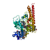
| |||||||||||||||||||||
|---|---|---|---|---|---|---|---|---|---|---|---|---|---|---|---|---|---|---|---|---|---|---|
| 1 | 
| |||||||||||||||||||||
| Unit cell |
| |||||||||||||||||||||
| Components on special symmetry positions |
|
-Components
-Protein , 1 types, 1 molecules Y
| #1: Protein | Mass: 55493.074 Da / Num. of mol.: 1 / Source method: isolated from a natural source / Source: (natural) Trichlorobacter ammonificans (bacteria) |
|---|
-Non-polymers , 5 types, 461 molecules 


| #2: Chemical | ChemComp-HEC /  Mass: 618.503 Da / Num. of mol.: 8 / Source method: obtained synthetically / Formula: C34H34FeN4O4 / Feature type: SUBJECT OF INVESTIGATION Mass: 618.503 Da / Num. of mol.: 8 / Source method: obtained synthetically / Formula: C34H34FeN4O4 / Feature type: SUBJECT OF INVESTIGATION#3: Chemical | ChemComp-NO / |  Mass: 30.006 Da / Num. of mol.: 1 / Source method: obtained synthetically / Formula: NO / Feature type: SUBJECT OF INVESTIGATION Mass: 30.006 Da / Num. of mol.: 1 / Source method: obtained synthetically / Formula: NO / Feature type: SUBJECT OF INVESTIGATION#4: Chemical | ChemComp-NO2 / |  Mass: 46.005 Da / Num. of mol.: 1 / Source method: obtained synthetically / Formula: NO2 / Feature type: SUBJECT OF INVESTIGATION Mass: 46.005 Da / Num. of mol.: 1 / Source method: obtained synthetically / Formula: NO2 / Feature type: SUBJECT OF INVESTIGATION#5: Chemical | ChemComp-CA / |  Mass: 40.078 Da / Num. of mol.: 1 / Source method: obtained synthetically / Formula: Ca / Feature type: SUBJECT OF INVESTIGATION Mass: 40.078 Da / Num. of mol.: 1 / Source method: obtained synthetically / Formula: Ca / Feature type: SUBJECT OF INVESTIGATION#6: Water | ChemComp-HOH / | Mass: 18.015 Da / Num. of mol.: 450 / Source method: isolated from a natural source / Formula: H2O |
|---|
-Details
| Has ligand of interest | Y |
|---|---|
| Has protein modification | Y |
-Experimental details
-Experiment
| Experiment | Method: X-RAY DIFFRACTION / Number of used crystals: 1 |
|---|
- Sample preparation
| Crystal | Density Matthews: 2.22 Å3/Da / Density % sol: 44.64 % |
|---|---|
| Crystal grow | Temperature: 293 K / Method: vapor diffusion, hanging drop Details: 20 mM NaNO2, 0.1 M Bis-Tris, pH 5.7, 17-20 % PEG 3350 |
-Data collection
| Diffraction | Mean temperature: 100 K / Serial crystal experiment: N |
|---|---|
| Diffraction source | Source: SYNCHROTRON / Site: ESRF  / Beamline: ID29 / Wavelength: 0.96862 Å / Beamline: ID29 / Wavelength: 0.96862 Å |
| Detector | Type: DECTRIS PILATUS 12M / Detector: PIXEL / Date: Sep 12, 2017 |
| Radiation | Protocol: SINGLE WAVELENGTH / Monochromatic (M) / Laue (L): M / Scattering type: x-ray |
| Radiation wavelength | Wavelength: 0.96862 Å / Relative weight: 1 |
| Reflection | Resolution: 1.55→49.6 Å / Num. obs: 69684 / % possible obs: 0.988 % / Redundancy: 4.4 % / CC1/2: 0.998 / Net I/σ(I): 11.4 |
| Reflection shell | Resolution: 1.55→1.59 Å / Num. unique obs: 5157 / CC1/2: 0.788 |
- Processing
| Software |
| ||||||||||||||||||||||||||||||||||||||||||||||||||||||||||||||||||||||||||||||||||||||||||||||||||||||||||||||||||||||||||||||||||||||||||||||||||||||||||||||||||||||||||||||||||||||
|---|---|---|---|---|---|---|---|---|---|---|---|---|---|---|---|---|---|---|---|---|---|---|---|---|---|---|---|---|---|---|---|---|---|---|---|---|---|---|---|---|---|---|---|---|---|---|---|---|---|---|---|---|---|---|---|---|---|---|---|---|---|---|---|---|---|---|---|---|---|---|---|---|---|---|---|---|---|---|---|---|---|---|---|---|---|---|---|---|---|---|---|---|---|---|---|---|---|---|---|---|---|---|---|---|---|---|---|---|---|---|---|---|---|---|---|---|---|---|---|---|---|---|---|---|---|---|---|---|---|---|---|---|---|---|---|---|---|---|---|---|---|---|---|---|---|---|---|---|---|---|---|---|---|---|---|---|---|---|---|---|---|---|---|---|---|---|---|---|---|---|---|---|---|---|---|---|---|---|---|---|---|---|---|
| Refinement | Method to determine structure: MOLECULAR REPLACEMENT / Resolution: 1.55→49.6 Å / Cor.coef. Fo:Fc: 0.964 / Cor.coef. Fo:Fc free: 0.942 / SU B: 3.768 / SU ML: 0.122 / Cross valid method: THROUGHOUT / ESU R: 0.101 / ESU R Free: 0.107 / Stereochemistry target values: MAXIMUM LIKELIHOOD
| ||||||||||||||||||||||||||||||||||||||||||||||||||||||||||||||||||||||||||||||||||||||||||||||||||||||||||||||||||||||||||||||||||||||||||||||||||||||||||||||||||||||||||||||||||||||
| Solvent computation | Ion probe radii: 0.8 Å / Shrinkage radii: 0.8 Å / VDW probe radii: 1.2 Å / Solvent model: BABINET MODEL WITH MASK | ||||||||||||||||||||||||||||||||||||||||||||||||||||||||||||||||||||||||||||||||||||||||||||||||||||||||||||||||||||||||||||||||||||||||||||||||||||||||||||||||||||||||||||||||||||||
| Displacement parameters | Biso mean: 25.172 Å2
| ||||||||||||||||||||||||||||||||||||||||||||||||||||||||||||||||||||||||||||||||||||||||||||||||||||||||||||||||||||||||||||||||||||||||||||||||||||||||||||||||||||||||||||||||||||||
| Refinement step | Cycle: 1 / Resolution: 1.55→49.6 Å
| ||||||||||||||||||||||||||||||||||||||||||||||||||||||||||||||||||||||||||||||||||||||||||||||||||||||||||||||||||||||||||||||||||||||||||||||||||||||||||||||||||||||||||||||||||||||
| Refine LS restraints |
|