 Movie
Movie Controller

[English] 日本語
 Yorodumi
- PDB-8rus: Hen egg-white lysozyme (HEWL) structure from EuXFEL FXE, multi-hi... -
+ Open data

- Basic information
| Entry | Database: PDB / ID: 8rus | ||||||
|---|---|---|---|---|---|---|---|
| Title | Hen egg-white lysozyme (HEWL) structure from EuXFEL FXE, multi-hit Droplet-on-Demand (DoD) injection, 9.3 keV photon energy, space group P432121 | ||||||
 Components Components | Lysozyme C | ||||||
 Keywords Keywords | HYDROLASE / XFEL / Lysozyme / HEWL / EuXFEL / European XFEL / Droplet on Demand / Droplet-on-Demand / DoD / multihit / multi-hit / kilohertz / SFX / Serial | ||||||
| Function / homology |  Function and homology information Function and homology informationLactose synthesis / Antimicrobial peptides / Neutrophil degranulation / beta-N-acetylglucosaminidase activity / cell wall macromolecule catabolic process / lysozyme / lysozyme activity / defense response to Gram-negative bacterium / killing of cells of another organism / defense response to Gram-positive bacterium ...Lactose synthesis / Antimicrobial peptides / Neutrophil degranulation / beta-N-acetylglucosaminidase activity / cell wall macromolecule catabolic process / lysozyme / lysozyme activity / defense response to Gram-negative bacterium / killing of cells of another organism / defense response to Gram-positive bacterium / defense response to bacterium / endoplasmic reticulum / extracellular space / identical protein binding / cytoplasm Similarity search - Function | ||||||
| Biological species |  | ||||||
| Method |  X-RAY DIFFRACTION / FREE ELECTRON LASER / MOLECULAR REPLACEMENT / Resolution: 1.38 Å X-RAY DIFFRACTION / FREE ELECTRON LASER / MOLECULAR REPLACEMENT / Resolution: 1.38 Å | ||||||
 Authors Authors | Perrett, S. / van Thor, J.J. | ||||||
| Funding support | European Union, 1items
| ||||||
 Citation Citation | Journal: Struct Dyn. / Year: 2024 Title: Kilohertz droplet-on-demand serial femtosecond crystallography at the European XFEL station FXE. Authors: Perrett, S. / Fadini, A. / Hutchison, C.D.M. / Bhattacharya, S. / Morrison, C. / Turkot, O. / Jakobsen, M.B. / Grossler, M. / Licon-Salaiz, J. / Griese, F. / Flewett, S. / Valerio, J. / ...Authors: Perrett, S. / Fadini, A. / Hutchison, C.D.M. / Bhattacharya, S. / Morrison, C. / Turkot, O. / Jakobsen, M.B. / Grossler, M. / Licon-Salaiz, J. / Griese, F. / Flewett, S. / Valerio, J. / Schulz, J. / Biednov, M. / Jiang, Y. / Han, H. / Yousef, H. / Khakhulin, D. / Milne, C. / Barty, A. / van Thor, J.J. | ||||||
| History |
|
- Structure visualization
| Structure viewer | Molecule: MolmilJmol/JSmol |
|---|
- Downloads & links
-Download
| PDBx/mmCIF format | 8rus.cif.gz | 93.5 KB | Display | PDBx/mmCIF format |
|---|---|---|---|---|
| PDB format | pdb8rus.ent.gz | 70.8 KB | Display | PDB format |
| PDBx/mmJSON format | 8rus.json.gz | Tree view | PDBx/mmJSON format | |
| Others |  Other downloads Other downloads |
-Validation report
| Summary document | 8rus_validation.pdf.gz | 436.5 KB | Display | wwPDB validaton report |
|---|---|---|---|---|
| Full document | 8rus_full_validation.pdf.gz | 436.6 KB | Display | |
| Data in XML | 8rus_validation.xml.gz | 8.4 KB | Display | |
| Data in CIF | 8rus_validation.cif.gz | 10.8 KB | Display | |
| Arichive directory | https://data.pdbj.org/pub/pdb/validation_reports/ru/8rusftp://data.pdbj.org/pub/pdb/validation_reports/ru/8rus | HTTPS FTP |
-Related structure data
| Similar structure data |
|---|
-Links
 PDBj
PDBj


- Assembly
| Deposited unit | 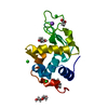
| ||||||||
|---|---|---|---|---|---|---|---|---|---|
| 1 |
| ||||||||
| Unit cell |
| ||||||||
| Components on special symmetry positions |
|
-Components
-Protein , 1 types, 1 molecules A
| #1: Protein | Mass: 14331.160 Da / Num. of mol.: 1 Source method: isolated from a genetically manipulated source Source: (gene. exp.)   Canavalia ensiformis (jack bean) / References: UniProt: P00698, lysozyme Canavalia ensiformis (jack bean) / References: UniProt: P00698, lysozyme |
|---|
-Non-polymers , 5 types, 86 molecules 


| #2: Chemical |  Mass: 35.453 Da / Num. of mol.: 3 / Source method: obtained synthetically / Formula: Cl Mass: 35.453 Da / Num. of mol.: 3 / Source method: obtained synthetically / Formula: Cl#3: Chemical | ChemComp-NA / |  Mass: 22.990 Da / Num. of mol.: 1 / Source method: obtained synthetically / Formula: Na Mass: 22.990 Da / Num. of mol.: 1 / Source method: obtained synthetically / Formula: Na#4: Chemical | ChemComp-ACT / |  Mass: 59.044 Da / Num. of mol.: 1 / Source method: obtained synthetically / Formula: C2H3O2 Mass: 59.044 Da / Num. of mol.: 1 / Source method: obtained synthetically / Formula: C2H3O2#5: Chemical |  Mass: 106.120 Da / Num. of mol.: 3 / Source method: obtained synthetically / Formula: C4H10O3 Mass: 106.120 Da / Num. of mol.: 3 / Source method: obtained synthetically / Formula: C4H10O3#6: Water | ChemComp-HOH / | Mass: 18.015 Da / Num. of mol.: 78 / Source method: isolated from a natural source / Formula: H2O |
|---|
-Details
| Has ligand of interest | N |
|---|---|
| Has protein modification | Y |
-Experimental details
-Experiment
| Experiment | Method: X-RAY DIFFRACTION / Number of used crystals: 1 |
|---|
- Sample preparation
| Crystal | Density Matthews: 2.11 Å3/Da / Density % sol: 41.73 % |
|---|---|
| Crystal grow | Temperature: 298 K / Method: batch mode / pH: 3.5 Details: 1:2 lysozyme (100 mg mL-1 in 50 mM NaOAc (pH 3.5)) and crystallization solution (0.1 M NaOAc (pH 3.5), 5% PEG6000 (v/v), 3.2 M NaCl) at 298K. |
-Data collection
| Diffraction | Mean temperature: 298 K / Serial crystal experiment: Y |
|---|---|
| Diffraction source | Source: FREE ELECTRON LASER / Site: European XFEL  / Beamline: FXE / Wavelength: 1.333 Å / Beamline: FXE / Wavelength: 1.333 Å |
| Detector | Type: STFC Large Pixel Detector / Detector: PIXEL / Date: Nov 2, 2022 |
| Radiation | Protocol: SINGLE WAVELENGTH / Monochromatic (M) / Laue (L): M / Scattering type: x-ray |
| Radiation wavelength | Wavelength: 1.333 Å / Relative weight: 1 |
| Reflection | Resolution: 1.38→18.73 Å / Num. obs: 12383860 / % possible obs: 100 % / Redundancy: 479 % / Biso Wilson estimate: 35.6 Å2 / CC1/2: 0.99 / CC star: 0.997 / R split: 0.0836 / Net I/σ(I): 6.77 |
| Reflection shell | Resolution: 1.38→1.4 Å / Redundancy: 43 % / Mean I/σ(I) obs: 0.9 / Num. unique obs: 107809 / CC1/2: 0.01 / CC star: 0.134 / R split: 0.5488 / % possible all: 100 |
| Serial crystallography measurement | Focal spot size: 10 µm2 / Pulse duration: 50 fsec. / Pulse photon energy: 9290 keV / XFEL pulse repetition rate: 47000 Hz |
| Serial crystallography sample delivery | Description: Droplet on Demand / Method: injection |
| Serial crystallography sample delivery injection | Carrier solvent: PEG / Crystal conc.: 20000000 / Description: Droplet on Demand Multi Hit / Filter size: 20 µm / Injector diameter: 80 µm / Injector temperature: 298 K / Power by: piezoelectric |
| Serial crystallography data reduction | Frames indexed: 74048 / Frames total: 255000 / XFEL run numbers: 3 |
- Processing
| Software |
| ||||||||||||||||||||||||||||||||||||||||||||||||||||||||||||||||||||||||||||||||||||||||||||||||||||||||||||||||||||||||||||||||||||||||||||
|---|---|---|---|---|---|---|---|---|---|---|---|---|---|---|---|---|---|---|---|---|---|---|---|---|---|---|---|---|---|---|---|---|---|---|---|---|---|---|---|---|---|---|---|---|---|---|---|---|---|---|---|---|---|---|---|---|---|---|---|---|---|---|---|---|---|---|---|---|---|---|---|---|---|---|---|---|---|---|---|---|---|---|---|---|---|---|---|---|---|---|---|---|---|---|---|---|---|---|---|---|---|---|---|---|---|---|---|---|---|---|---|---|---|---|---|---|---|---|---|---|---|---|---|---|---|---|---|---|---|---|---|---|---|---|---|---|---|---|---|---|---|
| Refinement | Method to determine structure: MOLECULAR REPLACEMENT / Resolution: 1.38→18.73 Å / SU ML: 0.34 / Cross valid method: FREE R-VALUE / σ(F): 1.33 / Phase error: 30.89 / Stereochemistry target values: ML
| ||||||||||||||||||||||||||||||||||||||||||||||||||||||||||||||||||||||||||||||||||||||||||||||||||||||||||||||||||||||||||||||||||||||||||||
| Solvent computation | Shrinkage radii: 0.9 Å / VDW probe radii: 1.1 Å / Solvent model: FLAT BULK SOLVENT MODEL | ||||||||||||||||||||||||||||||||||||||||||||||||||||||||||||||||||||||||||||||||||||||||||||||||||||||||||||||||||||||||||||||||||||||||||||
| Refinement step | Cycle: LAST / Resolution: 1.38→18.73 Å
| ||||||||||||||||||||||||||||||||||||||||||||||||||||||||||||||||||||||||||||||||||||||||||||||||||||||||||||||||||||||||||||||||||||||||||||
| Refine LS restraints |
| ||||||||||||||||||||||||||||||||||||||||||||||||||||||||||||||||||||||||||||||||||||||||||||||||||||||||||||||||||||||||||||||||||||||||||||
| LS refinement shell |
|
