[English] 日本語
Yorodumi
- PDB-8rk1: Crystal structure of FutA bound to Fe(III) solved by neutron diff... -

+
Open data


ID or keywords:

Loading...

-
Basic information

Entry
Database: PDB / ID: 8rk1
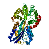
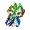
TitleCrystal structure of FutA bound to Fe(III) solved by neutron diffraction
ComponentsPutative iron ABC transporter, substrate binding protein
KeywordsMETAL BINDING PROTEIN / Periplasmic iron-binding protein / metal ion binding / iron homeostasis
Function / homologyBacterial extracellular solute-binding protein / Ferric binding protein / iron ion transport / metal ion binding / : / Putative iron ABC transporter, substrate binding protein
Function and homology information
Biological speciesProchlorococcus marinus subsp. pastoris str. CCMP1986 (bacteria)
MethodNEUTRON DIFFRACTION / NUCLEAR REACTOR / MOLECULAR REPLACEMENT / Resolution: 2.095 Å
AuthorsBolton, R. / Tews, I.
Funding support United Kingdom, 2items
OrganizationGrant numberCountry
University of Southampton United Kingdom
Diamond Light Source United Kingdom
Citation
Journal: Proc.Natl.Acad.Sci.USA / Year: 2024
Title: A redox switch allows binding of Fe(II) and Fe(III) ions in the cyanobacterial iron-binding protein FutA from Prochlorococcus.
Authors: Bolton, R. / Machelett, M.M. / Stubbs, J. / Axford, D. / Caramello, N. / Catapano, L. / Maly, M. / Rodrigues, M.J. / Cordery, C. / Tizzard, G.J. / MacMillan, F. / Engilberge, S. / von ...Authors: Bolton, R. / Machelett, M.M. / Stubbs, J. / Axford, D. / Caramello, N. / Catapano, L. / Maly, M. / Rodrigues, M.J. / Cordery, C. / Tizzard, G.J. / MacMillan, F. / Engilberge, S. / von Stetten, D. / Tosha, T. / Sugimoto, H. / Worrall, J.A.R. / Webb, J.S. / Zubkov, M. / Coles, S. / Mathieu, E. / Steiner, R.A. / Murshudov, G. / Schrader, T.E. / Orville, A.M. / Royant, A. / Evans, G. / Hough, M.A. / Owen, R.L. / Tews, I.
#1: Journal: Biorxiv / Year: 2023
Title: A redox switch allows binding of Fe(II) and Fe(III) ions in the cyanobacterial iron binding protein FutA from Prochlorococcus;
Authors: Bolton, R. / Machelett, M. / Stubbs, J. / Axford, D. / Caramello, N. / Catapano, L. / Maly, M. / Rodrigues, M. / Cordery, C. / Tizzard, G. / MacMillan, F. / Engilberge, S. / Stetten, D. / ...Authors: Bolton, R. / Machelett, M. / Stubbs, J. / Axford, D. / Caramello, N. / Catapano, L. / Maly, M. / Rodrigues, M. / Cordery, C. / Tizzard, G. / MacMillan, F. / Engilberge, S. / Stetten, D. / Tosha, T. / Sugimoto, H. / Worrall, J. / Webb, J. / Zubkov, M. / Coles, S. / Mathieu, E. / Steiner, R. / Murshudov, G. / Schrader, T. / Orville, A. / Royant, A. / Evans, G. / Hough, M. / Owen, R. / Tews, I.
History
DepositionDec 22, 2023Deposition site: PDBE / Processing site: PDBE
SupersessionJan 17, 2024ID: 8OEN
Revision 1.0Jan 17, 2024Provider: repository / Type: Initial release
Revision 1.1Mar 13, 2024Group: Database references / Category: citation / citation_author
Item: _citation.country / _citation.journal_abbrev ..._citation.country / _citation.journal_abbrev / _citation.journal_id_CSD / _citation.journal_id_ISSN / _citation.pdbx_database_id_DOI / _citation.title / _citation.year
Revision 1.2Mar 27, 2024Group: Database references / Category: citation / citation_author

-
Structure visualization

Structure viewerMolecule:
MolmilJmol/JSmol

Downloads & links

-
Assembly

Deposited unit
A: Putative iron ABC transporter, substrate binding protein
hetero molecules


Theoretical massNumber of molelcules
Total (without water)35,2752
Polymers35,2191
Non-polymers561
Water1,964109
1


  • Idetical with deposited unit
  • defined by author&software
  • Evidence: gel filtration
TypeNameSymmetry operationNumber
identity operation1_555x,y,z1
Buried area140 Å2
ΔGint-13 kcal/mol
Surface area13890 Å2
MethodPISA
Unit cell
Length a, b, c (Å)39.542, 78.316, 47.893
Angle α, β, γ (deg.)90, 97.365, 90
Int Tables number4
Space group name H-MP1211

-
Components

#1: Protein Putative iron ABC transporter, substrate binding protein


Mass: 35219.246 Da / Num. of mol.: 1
Source method: isolated from a genetically manipulated source
Source: (gene. exp.) Prochlorococcus marinus subsp. pastoris str. CCMP1986 (bacteria)
Gene: futA,sfuA, idiA, PMM1164 / Plasmid: pET- 24b(+) / Production host: Escherichia coli BL21(DE3) (bacteria) / References: UniProt: Q7V0T9
#2: Chemical ChemComp-FE / FE (III) ION / Iron


Mass: 55.845 Da / Num. of mol.: 1 / Source method: obtained synthetically / Formula: Fe / Feature type: SUBJECT OF INVESTIGATION
#3: Water ChemComp-HOH / water / Water


Mass: 18.015 Da / Num. of mol.: 109 / Source method: isolated from a natural source / Formula: H2O
Has ligand of interestY

-
Experimental details

-
Experiment

ExperimentMethod: NEUTRON DIFFRACTION / Number of used crystals: 1

-
Sample preparation

CrystalDensity Matthews: 2.09 Å3/Da / Density % sol: 41.22 %
Crystal growTemperature: 294 K / Method: batch mode
Details: Purified protein and crystallisation buffer were mixed at a 1:1 ratio. Crystallisation buffer was 20% PEG 3350, 200 mM sodium thiocyanate.

-
Data collection

DiffractionMean temperature: 294 K / Serial crystal experiment: N
Diffraction sourceSource: NUCLEAR REACTOR / Site: FRM II / Beamline: BIODIFF / Wavelength: 3.1 Å
DetectorType: BIODIFF / Detector: IMAGE PLATE / Date: Feb 2, 2020
RadiationProtocol: SINGLE WAVELENGTH / Monochromatic (M) / Laue (L): M / Scattering type: neutron
Radiation wavelengthWavelength: 3.1 Å / Relative weight: 1
ReflectionResolution: 2.095→50 Å / Num. obs: 13758 / % possible obs: 80.8 % / Redundancy: 1.9 % / CC1/2: 0.971 / Rpim(I) all: 0.106 / Rrim(I) all: 0.168 / Net I/σ(I): 4.6
Reflection shellResolution: 2.095→2.18 Å / Num. unique obs: 886 / CC1/2: 0.716 / Rpim(I) all: 0.334 / Rrim(I) all: 0.473 / % possible all: 52.4

-
Processing

RefinementMethod to determine structure: MOLECULAR REPLACEMENT / Resolution: 2.095→24.95 Å / Cor.coef. Fo:Fc: 0.936 / Cor.coef. Fo:Fc free: 0.869 / WRfactor Rfree: 0.243 / WRfactor Rwork: 0.176 / SU B: 10.369 / SU ML: 0.267 / Average fsc free: 0.9545 / Average fsc work: 0.976 / Cross valid method: FREE R-VALUE / ESU R Free: 0.357
Details: Hydrogens have been used if present in the input file
RfactorNum. reflection% reflection
Rfree0.2425 698 5.077 %
Rwork0.1776 13050 -
all0.181 --
obs-13748 80.412 %
Solvent computationIon probe radii: 0.8 Å / Shrinkage radii: 0.8 Å / VDW probe radii: 1.2 Å / Solvent model: MASK BULK SOLVENT
Displacement parametersBiso mean: 22.465 Å2
Baniso -1Baniso -2Baniso -3
1--0.134 Å2-0 Å2-0.184 Å2
2--0.197 Å20 Å2
3----0.014 Å2
Refine LS restraints
Refine-IDTypeDev idealDev ideal targetNumber
NEUTRON DIFFRACTIONr_bond_refined_d0.0280.0145311
NEUTRON DIFFRACTIONr_angle_refined_deg0.951.6319360
NEUTRON DIFFRACTIONr_dihedral_angle_1_deg6.3575319
NEUTRON DIFFRACTIONr_dihedral_angle_2_deg7.6577.05690
NEUTRON DIFFRACTIONr_dihedral_angle_3_deg14.61910735
NEUTRON DIFFRACTIONr_dihedral_angle_6_deg14.92610136
NEUTRON DIFFRACTIONr_chiral_restr0.0610.2382
NEUTRON DIFFRACTIONr_gen_planes_refined0.0060.023591
NEUTRON DIFFRACTIONr_nbd_refined0.190.22136
NEUTRON DIFFRACTIONr_nbtor_refined0.1630.21384
NEUTRON DIFFRACTIONr_xyhbond_nbd_refined0.2030.2130
NEUTRON DIFFRACTIONr_metal_ion_refined0.0220.22
NEUTRON DIFFRACTIONr_symmetry_nbd_refined0.2260.250
NEUTRON DIFFRACTIONr_symmetry_xyhbond_nbd_refined0.2270.212
NEUTRON DIFFRACTIONr_mcbond_it1.0211.9011867
NEUTRON DIFFRACTIONr_mcangle_it1.7693.2352792
NEUTRON DIFFRACTIONr_scbond_it1.2832.3523443
NEUTRON DIFFRACTIONr_scangle_it2.3223.6616567
NEUTRON DIFFRACTIONr_lrange_it6.80322.7585806
LS refinement shell

Refine-ID: NEUTRON DIFFRACTION / Total num. of bins used: 20

Resolution (Å)Rfactor RfreeNum. reflection RfreeRfactor RworkNum. reflection RworkRfactor allNum. reflection allFsc freeFsc work% reflection obs (%)WRfactor Rwork
2.095-2.1490.33290.2995560.312440.9210.94447.02570.287
2.149-2.2070.4310.2656730.26912210.8910.95657.65770.264
2.207-2.2710.282480.2427350.24411890.9540.96265.85370.243
2.271-2.340.259420.2178670.21911680.9560.96877.82530.207
2.34-2.4160.236500.1868360.18911110.9650.97779.7480.179
2.416-2.50.232480.1838550.18510920.9670.97882.69230.174
2.5-2.5930.284530.2087790.21310120.940.96982.21340.213
2.593-2.6980.319390.2177480.22110090.920.96877.9980.225
2.698-2.8170.254490.2088090.2119760.9530.97187.90980.215
2.817-2.9520.168340.177960.179100.9750.98291.20880.164
2.952-3.1090.291410.177760.1778830.9590.98292.52550.166
3.109-3.2950.213330.1657670.1678440.9670.98494.78670.164
3.295-3.5180.205360.1527090.1557790.970.98795.63540.154
3.518-3.7940.303290.1496680.1567320.9510.98695.21860.156
3.794-4.1460.235370.1446080.1496840.9570.98894.29820.155
4.146-4.620.207270.1245360.1286110.9740.99192.1440.14
4.62-5.3040.199260.1344750.1375510.9760.9990.92560.149
5.304-6.4240.207290.1543810.1574670.9720.98687.79440.181
6.424-8.7980.10490.1233000.1223650.9940.99184.65750.154
8.798-24.950.14380.1841760.1822380.9840.98577.31090.242

+
About Yorodumi

-
News

-
Feb 9, 2022. New format data for meta-information of EMDB entries

New format data for meta-information of EMDB entries

  • Version 3 of the EMDB header file is now the official format.
  • The previous official version 1.9 will be removed from the archive.

Related info.:EMDB header

External links:wwPDB to switch to version 3 of the EMDB data model

-
Aug 12, 2020. Covid-19 info

Covid-19 info

URL: https://pdbj.org/emnavi/covid19.php

New page: Covid-19 featured information page in EM Navigator.

Related info.:Covid-19 info / Mar 5, 2020. Novel coronavirus structure data

+
Mar 5, 2020. Novel coronavirus structure data

Novel coronavirus structure data

Related info.:Yorodumi Speices / Aug 12, 2020. Covid-19 info

External links:COVID-19 featured content - PDBj / Molecule of the Month (242):Coronavirus Proteases

+
Jan 31, 2019. EMDB accession codes are about to change! (news from PDBe EMDB page)

EMDB accession codes are about to change! (news from PDBe EMDB page)

  • The allocation of 4 digits for EMDB accession codes will soon come to an end. Whilst these codes will remain in use, new EMDB accession codes will include an additional digit and will expand incrementally as the available range of codes is exhausted. The current 4-digit format prefixed with “EMD-” (i.e. EMD-XXXX) will advance to a 5-digit format (i.e. EMD-XXXXX), and so on. It is currently estimated that the 4-digit codes will be depleted around Spring 2019, at which point the 5-digit format will come into force.
  • The EM Navigator/Yorodumi systems omit the EMD- prefix.

Related info.:Q: What is EMD? / ID/Accession-code notation in Yorodumi/EM Navigator

External links:EMDB Accession Codes are Changing Soon! / Contact to PDBj

+
Jul 12, 2017. Major update of PDB

Major update of PDB

  • wwPDB released updated PDB data conforming to the new PDBx/mmCIF dictionary.
  • This is a major update changing the version number from 4 to 5, and with Remediation, in which all the entries are updated.
  • In this update, many items about electron microscopy experimental information are reorganized (e.g. em_software).
  • Now, EM Navigator and Yorodumi are based on the updated data.

External links:wwPDB Remediation / Enriched Model Files Conforming to OneDep Data Standards Now Available in the PDB FTP Archive

-
Yorodumi

Thousand views of thousand structures

  • Yorodumi is a browser for structure data from EMDB, PDB, SASBDB, etc.
  • This page is also the successor to EM Navigator detail page, and also detail information page/front-end page for Omokage search.
  • The word "yorodu" (or yorozu) is an old Japanese word meaning "ten thousand". "mi" (miru) is to see.

Related info.:EMDB / PDB / SASBDB / Comparison of 3 databanks / Yorodumi Search / Aug 31, 2016. New EM Navigator & Yorodumi / Yorodumi Papers / Jmol/JSmol / Function and homology information / Changes in new EM Navigator and Yorodumi

Read more