 Movie
Movie Controller

[English] 日本語
 Yorodumi
- PDB-8rjs: Serial femtosecond X-ray structure of a fluorescence optimized ba... -
+ Open data

- Basic information
| Entry | Database: PDB / ID: 8rjs | ||||||||||||||||||||||||||||||
|---|---|---|---|---|---|---|---|---|---|---|---|---|---|---|---|---|---|---|---|---|---|---|---|---|---|---|---|---|---|---|---|
| Title | Serial femtosecond X-ray structure of a fluorescence optimized bathy phytochrome PAiRFP2 derived from wild-type Agp2 in I5 intermediate state. | ||||||||||||||||||||||||||||||
 Components Components | histidine kinase | ||||||||||||||||||||||||||||||
 Keywords Keywords | SIGNALING PROTEIN / fluorescent protein / phytochrome / bacteriophytochrome / intermediate / optogenetic | ||||||||||||||||||||||||||||||
| Function / homology |  Function and homology information Function and homology informationprotein histidine kinase activity / detection of visible light / histidine kinase / phosphorelay signal transduction system / photoreceptor activity / regulation of DNA-templated transcription / ATP binding Similarity search - Function | ||||||||||||||||||||||||||||||
| Biological species |  Agrobacterium fabrum str. C58 (bacteria) Agrobacterium fabrum str. C58 (bacteria) | ||||||||||||||||||||||||||||||
| Method |  X-RAY DIFFRACTION / FREE ELECTRON LASER / MOLECULAR REPLACEMENT / Resolution: 2.43 Å X-RAY DIFFRACTION / FREE ELECTRON LASER / MOLECULAR REPLACEMENT / Resolution: 2.43 Å | ||||||||||||||||||||||||||||||
 Authors Authors | Sauthof, L. / Schmidt, A. / Szczepek, M. / Brewster, A.S. / Kern, J.F. / Scheerer, P. | ||||||||||||||||||||||||||||||
| Funding support |  Germany, Germany,  Israel, Israel,  United States, United States,  United Kingdom, 9items United Kingdom, 9items
| ||||||||||||||||||||||||||||||
 Citation Citation | Journal: Sci Adv / Year: 2025 Title: Serial-femtosecond crystallography reveals how a phytochrome variant couples chromophore and protein structural changes. Authors: Sauthof, L. / Szczepek, M. / Schmidt, A. / Bhowmick, A. / Dasgupta, M. / Mackintosh, M.J. / Gul, S. / Fuller, F.D. / Chatterjee, R. / Young, I.D. / Michael, N. / Heyder, N.A. / Bauer, B. / ...Authors: Sauthof, L. / Szczepek, M. / Schmidt, A. / Bhowmick, A. / Dasgupta, M. / Mackintosh, M.J. / Gul, S. / Fuller, F.D. / Chatterjee, R. / Young, I.D. / Michael, N. / Heyder, N.A. / Bauer, B. / Koch, A. / Bogacz, I. / Kim, I.S. / Simon, P.S. / Butryn, A. / Aller, P. / Chukhutsina, V.U. / Baxter, J.M. / Hutchison, C.D.M. / Liebschner, D. / Poon, B. / Sauter, N.K. / Miller, M.D. / Alonso-Mori, R. / Hunter, M.S. / Batyuk, A. / Owada, S. / Tono, K. / Tanaka, R. / van Thor, J.J. / Krauss, N. / Lamparter, T. / Brewster, A.S. / Schapiro, I. / Orville, A.M. / Yachandra, V.K. / Yano, J. / Hildebrandt, P. / Kern, J.F. / Scheerer, P. | ||||||||||||||||||||||||||||||
| History |
|
- Structure visualization
| Structure viewer | Molecule: MolmilJmol/JSmol |
|---|
- Downloads & links
-Download
| PDBx/mmCIF format | 8rjs.cif.gz | 207.9 KB | Display | PDBx/mmCIF format |
|---|---|---|---|---|
| PDB format | pdb8rjs.ent.gz | 164.2 KB | Display | PDB format |
| PDBx/mmJSON format | 8rjs.json.gz | Tree view | PDBx/mmJSON format | |
| Others |  Other downloads Other downloads |
-Validation report
| Summary document | 8rjs_validation.pdf.gz | 1.9 MB | Display | wwPDB validaton report |
|---|---|---|---|---|
| Full document | 8rjs_full_validation.pdf.gz | 1.9 MB | Display | |
| Data in XML | 8rjs_validation.xml.gz | 43.5 KB | Display | |
| Data in CIF | 8rjs_validation.cif.gz | 56.2 KB | Display | |
| Arichive directory | https://data.pdbj.org/pub/pdb/validation_reports/rj/8rjsftp://data.pdbj.org/pub/pdb/validation_reports/rj/8rjs | HTTPS FTP |
-Related structure data
| Related structure data |  8rjmC  8rjnC  8rjoC  8rjpC  8rjqC  8rjrC  8rjtC  8rjuC C: citing same article ( |
|---|---|
| Similar structure data |


-Links
 PDBj
PDBj


- Assembly
| Deposited unit | 
| |||||||||||||||
|---|---|---|---|---|---|---|---|---|---|---|---|---|---|---|---|---|
| 1 | 
| |||||||||||||||
| 2 | 
| |||||||||||||||
| Unit cell |
| |||||||||||||||
| Components on special symmetry positions |
|
-Components
| #1: Protein | Mass: 57507.484 Da / Num. of mol.: 2 Mutation: K69R, R83K, G120D, A123T, M163L, Q168E, R220P, S243N, V244F, G269D, A276V, Y270C, E294A, H303F, H333R, I336L, D349R, M351I, A386V, G409D, L419I, T469S, A487T, E494G Source method: isolated from a genetically manipulated source Source: (gene. exp.) Agrobacterium fabrum str. C58 (bacteria)Gene: Atu2165 / Production host: #2: Chemical | 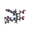  Mass: 584.662 Da / Num. of mol.: 2 / Source method: obtained synthetically / Formula: C33H36N4O6 / Feature type: SUBJECT OF INVESTIGATION Mass: 584.662 Da / Num. of mol.: 2 / Source method: obtained synthetically / Formula: C33H36N4O6 / Feature type: SUBJECT OF INVESTIGATION#3: Chemical | ChemComp-SO4 /   Mass: 96.063 Da / Num. of mol.: 5 / Source method: obtained synthetically / Formula: SO4 Mass: 96.063 Da / Num. of mol.: 5 / Source method: obtained synthetically / Formula: SO4#4: Chemical | ChemComp-CL / |   Mass: 35.453 Da / Num. of mol.: 1 / Source method: obtained synthetically / Formula: Cl Mass: 35.453 Da / Num. of mol.: 1 / Source method: obtained synthetically / Formula: Cl#5: Water | ChemComp-HOH / |  Mass: 18.015 Da / Num. of mol.: 88 / Source method: isolated from a natural source / Formula: H2O Mass: 18.015 Da / Num. of mol.: 88 / Source method: isolated from a natural source / Formula: H2OHas ligand of interest | Y | Has protein modification | Y | |
|---|
-Experimental details
-Experiment
| Experiment | Method: X-RAY DIFFRACTION / Number of used crystals: 1 |
|---|
- Sample preparation
| Crystal | Density Matthews: 3.9 Å3/Da / Density % sol: 68.5 % |
|---|---|
| Crystal grow | Temperature: 279 K / Method: batch mode Details: 1.0 - 2.2 M ammonium sulphate, 2 - 12% PEG 1000, 0.1 M HEPES pH 6.8 - 7.7, 0.025% low-melt agarose |
-Data collection
| Diffraction | Mean temperature: 298 K / Serial crystal experiment: N |
|---|---|
| Diffraction source | Source: FREE ELECTRON LASER / Site: SACLA  / Beamline: BL2 / Wavelength: 1.24028 Å / Beamline: BL2 / Wavelength: 1.24028 Å |
| Detector | Type: MPCCD / Detector: CCD / Date: Feb 17, 2019 |
| Radiation | Protocol: SINGLE WAVELENGTH / Monochromatic (M) / Laue (L): M / Scattering type: x-ray |
| Radiation wavelength | Wavelength: 1.24028 Å / Relative weight: 1 |
| Reflection | Resolution: 2.43→92.22 Å / Num. obs: 68292 / % possible obs: 100 % / Redundancy: 194.9 % / CC1/2: 0.978 / Net I/σ(I): 19.8 |
| Reflection shell | Resolution: 2.43→2.49 Å / Num. unique obs: 4822 / CC1/2: 0.014 |
- Processing
| Software |
| ||||||||||||||||||||||||||||||||||||||||||||||||||||||||||||||||||||||||||||||||||||||||||||||||||||||||||||||||||||||||||||||||||||||||||||||||||||||||||||||||||||||||||||||||||||||
|---|---|---|---|---|---|---|---|---|---|---|---|---|---|---|---|---|---|---|---|---|---|---|---|---|---|---|---|---|---|---|---|---|---|---|---|---|---|---|---|---|---|---|---|---|---|---|---|---|---|---|---|---|---|---|---|---|---|---|---|---|---|---|---|---|---|---|---|---|---|---|---|---|---|---|---|---|---|---|---|---|---|---|---|---|---|---|---|---|---|---|---|---|---|---|---|---|---|---|---|---|---|---|---|---|---|---|---|---|---|---|---|---|---|---|---|---|---|---|---|---|---|---|---|---|---|---|---|---|---|---|---|---|---|---|---|---|---|---|---|---|---|---|---|---|---|---|---|---|---|---|---|---|---|---|---|---|---|---|---|---|---|---|---|---|---|---|---|---|---|---|---|---|---|---|---|---|---|---|---|---|---|---|---|
| Refinement | Method to determine structure: MOLECULAR REPLACEMENT / Resolution: 2.43→92.22 Å / Cor.coef. Fo:Fc: 0.962 / Cor.coef. Fo:Fc free: 0.944 / SU B: 11.88 / SU ML: 0.235 / Cross valid method: THROUGHOUT / ESU R: 0.243 / ESU R Free: 0.209 / Stereochemistry target values: MAXIMUM LIKELIHOOD / Details: HYDROGENS HAVE BEEN ADDED IN THE RIDING POSITIONS
| ||||||||||||||||||||||||||||||||||||||||||||||||||||||||||||||||||||||||||||||||||||||||||||||||||||||||||||||||||||||||||||||||||||||||||||||||||||||||||||||||||||||||||||||||||||||
| Solvent computation | Ion probe radii: 0.8 Å / Shrinkage radii: 0.8 Å / VDW probe radii: 1.2 Å / Solvent model: MASK | ||||||||||||||||||||||||||||||||||||||||||||||||||||||||||||||||||||||||||||||||||||||||||||||||||||||||||||||||||||||||||||||||||||||||||||||||||||||||||||||||||||||||||||||||||||||
| Displacement parameters | Biso mean: 69.59 Å2
| ||||||||||||||||||||||||||||||||||||||||||||||||||||||||||||||||||||||||||||||||||||||||||||||||||||||||||||||||||||||||||||||||||||||||||||||||||||||||||||||||||||||||||||||||||||||
| Refinement step | Cycle: 1 / Resolution: 2.43→92.22 Å
| ||||||||||||||||||||||||||||||||||||||||||||||||||||||||||||||||||||||||||||||||||||||||||||||||||||||||||||||||||||||||||||||||||||||||||||||||||||||||||||||||||||||||||||||||||||||
| Refine LS restraints |
|