 Movie
Movie Controller


+ Open data

- Basic information
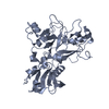
| Entry | Database: PDB / ID: 8rek | ||||||
|---|---|---|---|---|---|---|---|
| Title | Plasmodium vivax Apical Membrane Antigen 1/Fab complex | ||||||
 Components Components |
| ||||||
 Keywords Keywords | IMMUNE SYSTEM / AMA-1 / Plasmodium vivax / Antigen / Fab | ||||||
| Function / homology | Apical membrane antigen 1 domain superfamily / Apical membrane antigen 1 / Apical membrane antigen 1 / Apical membrane antigen 1 / membrane / Apical membrane antigen 1 Function and homology information Function and homology information | ||||||
| Biological species |   | ||||||
| Method |  X-RAY DIFFRACTION / SYNCHROTRON / MOLECULAR REPLACEMENT / Resolution: 3.05 Å X-RAY DIFFRACTION / SYNCHROTRON / MOLECULAR REPLACEMENT / Resolution: 3.05 Å | ||||||
 Authors Authors | Bentley, G.A. / Saul, F.A. / Vulliez-LeNormand, B. | ||||||
| Funding support |  France, 1items France, 1items
| ||||||
 Citation Citation | Journal: J Struct Biol X / Year: 2024 Title: Conformational variability in the D2 loop of Plasmodium Apical Membrane antigen 1. Authors: Saul, F.A. / Vulliez-Le Normand, B. / Boes, A. / Spiegel, H. / Kocken, C.H.M. / Faber, B.W. / Bentley, G.A. | ||||||
| History |
|
- Structure visualization
| Structure viewer | Molecule: MolmilJmol/JSmol |
|---|
- Downloads & links
-Download
| PDBx/mmCIF format | 8rek.cif.gz | 339.3 KB | Display | PDBx/mmCIF format |
|---|---|---|---|---|
| PDB format | pdb8rek.ent.gz | 273 KB | Display | PDB format |
| PDBx/mmJSON format | 8rek.json.gz | Tree view | PDBx/mmJSON format | |
| Others |  Other downloads Other downloads |
-Validation report
| Summary document | 8rek_validation.pdf.gz | 449.2 KB | Display | wwPDB validaton report |
|---|---|---|---|---|
| Full document | 8rek_full_validation.pdf.gz | 449.9 KB | Display | |
| Data in XML | 8rek_validation.xml.gz | 31.2 KB | Display | |
| Data in CIF | 8rek_validation.cif.gz | 41.6 KB | Display | |
| Arichive directory | https://data.pdbj.org/pub/pdb/validation_reports/re/8rekftp://data.pdbj.org/pub/pdb/validation_reports/re/8rek | HTTPS FTP |
-Related structure data


-Links
 PDBj
PDBj

- Assembly
| Deposited unit | 
| ||||||||
|---|---|---|---|---|---|---|---|---|---|
| 1 |
| ||||||||
| Unit cell |
|
-Components
| #1: Protein | Mass: 54180.820 Da / Num. of mol.: 1 Mutation: S178N, N226D, N441Q are potential N-glycosylation sites Source method: isolated from a genetically manipulated source Source: (gene. exp.)  Komagataella pastoris (fungus) / References: UniProt: Q9TY14 Komagataella pastoris (fungus) / References: UniProt: Q9TY14 |
|---|---|
| #2: Antibody | Mass: 23539.311 Da / Num. of mol.: 1 / Source method: isolated from a natural source / Source: (natural) |
| #3: Antibody | Mass: 23296.701 Da / Num. of mol.: 1 / Source method: isolated from a natural source / Source: (natural) |
| Has ligand of interest | Y |
| Has protein modification | Y |
-Experimental details
-Experiment
| Experiment | Method: X-RAY DIFFRACTION / Number of used crystals: 1 |
|---|
- Sample preparation
| Crystal | Density Matthews: 2.2 Å3/Da / Density % sol: 44 % |
|---|---|
| Crystal grow | Temperature: 290 K / Method: vapor diffusion, hanging drop / pH: 7 Details: Crystals were obtained by mixing 0.8 micro-L of PvAMA1-Fab complex with 0.2 micro-L of 15% 1,2,3 heptanetriol and 0.8 micro-L of reservoir buffer comprising 20% PEG 2000 monomethylether and ...Details: Crystals were obtained by mixing 0.8 micro-L of PvAMA1-Fab complex with 0.2 micro-L of 15% 1,2,3 heptanetriol and 0.8 micro-L of reservoir buffer comprising 20% PEG 2000 monomethylether and 0.1 M Tris pH 7. The final protein concentration was 3.2 mg/ml. |
-Data collection
| Diffraction | Mean temperature: 100 K / Serial crystal experiment: N |
|---|---|
| Diffraction source | Source: SYNCHROTRON / Site: ESRF / Beamline: ID14-1 / Wavelength: 0.934 Å |
| Detector | Type: ADSC QUANTUM 210r / Detector: CCD / Date: Sep 27, 2006 |
| Radiation | Protocol: SINGLE WAVELENGTH / Monochromatic (M) / Laue (L): M / Scattering type: x-ray |
| Radiation wavelength | Wavelength: 0.934 Å / Relative weight: 1 |
| Reflection | Resolution: 3.05→73.39 Å / Num. obs: 18062 / % possible obs: 99.9 % / Redundancy: 7.1 % / CC1/2: 0.996 / Rmerge(I) obs: 0.167 / Rpim(I) all: 0.067 / Rrim(I) all: 0.181 / Χ2: 0.97 / Net I/σ(I): 11.8 / Num. measured all: 128197 |
| Reflection shell | Resolution: 3.05→3.26 Å / % possible obs: 100 % / Redundancy: 7.2 % / Rmerge(I) obs: 1.203 / Num. measured all: 23088 / Num. unique obs: 3193 / CC1/2: 0.316 / Rpim(I) all: 0.48 / Rrim(I) all: 1.296 / Χ2: 0.94 / Net I/σ(I) obs: 1.7 |
- Processing
| Software |
| ||||||||||||||||||||||||||||||||||||||||||||||||||||||||||||||||||||||||||||||||||||||||||||||||||||||||||||||||||||||||||||||||||||||||||||||||||||||||||||||||||||||||||||||||||||||
|---|---|---|---|---|---|---|---|---|---|---|---|---|---|---|---|---|---|---|---|---|---|---|---|---|---|---|---|---|---|---|---|---|---|---|---|---|---|---|---|---|---|---|---|---|---|---|---|---|---|---|---|---|---|---|---|---|---|---|---|---|---|---|---|---|---|---|---|---|---|---|---|---|---|---|---|---|---|---|---|---|---|---|---|---|---|---|---|---|---|---|---|---|---|---|---|---|---|---|---|---|---|---|---|---|---|---|---|---|---|---|---|---|---|---|---|---|---|---|---|---|---|---|---|---|---|---|---|---|---|---|---|---|---|---|---|---|---|---|---|---|---|---|---|---|---|---|---|---|---|---|---|---|---|---|---|---|---|---|---|---|---|---|---|---|---|---|---|---|---|---|---|---|---|---|---|---|---|---|---|---|---|---|---|
| Refinement | Method to determine structure: MOLECULAR REPLACEMENT / Resolution: 3.05→69.54 Å / Cor.coef. Fo:Fc: 0.912 / Cor.coef. Fo:Fc free: 0.866 / SU B: 68.08 / SU ML: 0.513 / Cross valid method: THROUGHOUT / ESU R Free: 0.551 / Stereochemistry target values: MAXIMUM LIKELIHOOD / Details: HYDROGENS HAVE BEEN ADDED IN THE RIDING POSITIONS
| ||||||||||||||||||||||||||||||||||||||||||||||||||||||||||||||||||||||||||||||||||||||||||||||||||||||||||||||||||||||||||||||||||||||||||||||||||||||||||||||||||||||||||||||||||||||
| Solvent computation | Ion probe radii: 0.8 Å / Shrinkage radii: 0.8 Å / VDW probe radii: 1.2 Å / Solvent model: MASK | ||||||||||||||||||||||||||||||||||||||||||||||||||||||||||||||||||||||||||||||||||||||||||||||||||||||||||||||||||||||||||||||||||||||||||||||||||||||||||||||||||||||||||||||||||||||
| Displacement parameters | Biso mean: 95.223 Å2
| ||||||||||||||||||||||||||||||||||||||||||||||||||||||||||||||||||||||||||||||||||||||||||||||||||||||||||||||||||||||||||||||||||||||||||||||||||||||||||||||||||||||||||||||||||||||
| Refinement step | Cycle: 1 / Resolution: 3.05→69.54 Å
| ||||||||||||||||||||||||||||||||||||||||||||||||||||||||||||||||||||||||||||||||||||||||||||||||||||||||||||||||||||||||||||||||||||||||||||||||||||||||||||||||||||||||||||||||||||||
| Refine LS restraints |
|