Entry Database : PDB / ID : 8raiTitle Crystal structure of D-amino acid transaminase from Haliscomenobacter hydrossis point mutant R90I complexed with phenylhydrazine Aminotransferase class IV Keywords / / / / / / / Function / homology Function Domain/homology Component
/ / / / / / / / / Biological species Haliscomenobacter hydrossis DSM 1100 (bacteria)Method / / Resolution : 2 Å Authors Matyuta, I.O. / Bakunova, A.K. / Minyaev, M.E. / Popov, V.O. / Bezsudnova, E.Y. / Boyko, K.M. Funding support Organization Grant number Country Russian Science Foundation 23-74-30004
Journal : Arch.Biochem.Biophys. / Year : 2024Title : Multifunctionality of arginine residues in the active sites of non-canonical d-amino acid transaminases.Authors : Bakunova, A.K. / Matyuta, I.O. / Minyaev, M.E. / Isaikina, T.Y. / Boyko, K.M. / Popov, V.O. / Bezsudnova, E.Y. History Deposition Dec 1, 2023 Deposition site / Processing site Revision 1.0 Dec 27, 2023 Provider / Type Revision 1.1 May 1, 2024 Group / Category / citation_authorItem _citation.country / _citation.journal_abbrev ... _citation.country / _citation.journal_abbrev / _citation.journal_id_ASTM / _citation.journal_id_CSD / _citation.journal_id_ISSN / _citation.page_first / _citation.page_last / _citation.pdbx_database_id_DOI / _citation.pdbx_database_id_PubMed / _citation.title / _citation.year / _citation_author.identifier_ORCID Revision 1.2 May 8, 2024 Group / Category / Item / _citation.title
Show all Show less
 Movie
Movie Controller

 Yorodumi
 Open data
 Basic information
 Components
 Keywords
 Function and homology information
 Haliscomenobacter hydrossis DSM 1100 (bacteria)
 X-RAY DIFFRACTION /
  Authors
 Russian Federation, 1items
  Citation
 Structure visualization
 Downloads & links
 Other downloads
 PDBj
PDBj
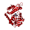
 Assembly
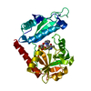
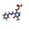
 Mass: 337.268 Da / Num. of mol.: 1 / Source method: obtained synthetically / Formula: C14H16N3O5P / Feature type: SUBJECT OF INVESTIGATION
 Mass: 92.094 Da / Num. of mol.: 1 / Source method: obtained synthetically / Formula: C3H8O3
 Mass: 18.015 Da / Num. of mol.: 205 / Source method: isolated from a natural source / Formula: H2O
 Sample preparation
 Processing